Alzheimer's Research - Ai-Peptides

FREE SHIPPING ON ORDERS OVER $100

Research Categories
[list_research_categories]
Research Categories

Alzheimer's Research

ALL ARTICLES AND PRODUCT INFORMATION PROVIDED ON THIS WEBSITE ARE FOR INFORMATIONAL AND EDUCATIONAL PURPOSES ONLY. The products offered on this website are furnished for in-vitro studies only. In-vitro studies (Latin: in glass) are performed outside of the body. These products are not medicines or drugs and have not been approved by the FDA to prevent, treat or cure any medical condition, ailment or disease. Bodily introduction of any kind into humans or animals is strictly forbidden by law.

The Role of Cell-Permeable Peptides and The JNK Family in Preventing Neuronal Degeneration

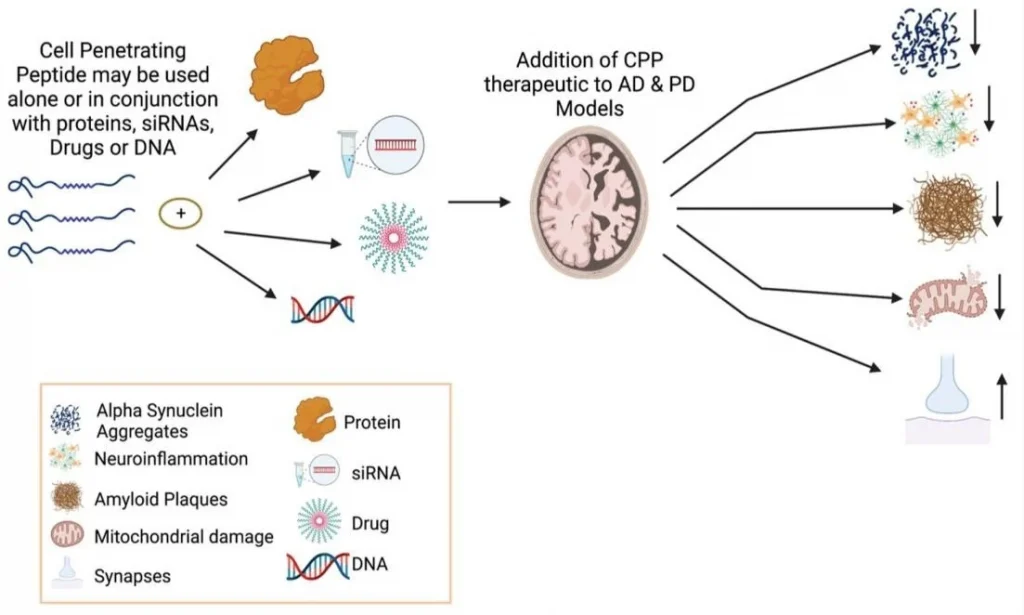
Figure 1:     Neuroprotective features of CPPs in combination with other molecules or peptides for treating PD and AD.

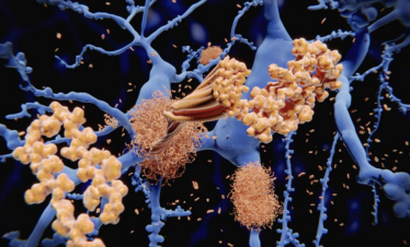
JNK family participates in the apoptosis pathway, also known as programmed cell death.[4] The apoptosis mechanism is used to eliminate cells that are damaged in anyway. This mechanism is suitable for healthy people because it helps avoid developing cancer and other disorders.[1] On the other hand, apoptosis is detrimental in neurodegenerative diseases that share the abnormal accumulation of misfolded proteins, contributing to dementia, cognitive loss, memory loss, behavioral problems, and sleep problems, among others, through neuronal cell loss.[3,5,7] In addition, the JNK family also participates in pathways related to regulation, plasticity, development of the Central Nervous System (CNS), inflammation, and autophagy.[9] The well-functioning of all these processes is vital. Recent studies suggested that an imbalance of the JNK family members can accelerate the progression of both AD and PD pathologies. One of the hallmarks of AD is the aggregation of amyloid beta (Aβ) in the extracellular area.[7] The overexpression of Aβ triggers the activation of JNK-3, which also participates in the formation of Aβ42, a toxic species that affects the neuron’s cell function when it accumulates.[7] It is unclear whether the neurodegenerative disorder or the JNK imbalance comes first. However, they both have a direct relationship where PD and AD patients show high levels of JNK in the brain (postmortem), and irregular levels of the JNK family accelerate the progression of both disorders (see Fig. 2). [5,9] JNK imbalance could also lead to increased inflammation responses, low plasticity, and a flaw in autophagy performance, among other related adverse effects. An increase in apoptosis in PD and AD patients causes a decrease in neurons in the brain, affecting cognitive, behavioral, and memory performance.[8] JNK also decreases the α-syn accumulation in PD models, contributing to neuroprotection.[10] JNK pathway influence positively many vital processes of the body unless an imbalance occurs. [2,8] JNK has been the target for treating several diseases like cancer, strokes, PD, AD, Huntington’s disease, and other neurodegenerative disorders, showing promising results. More investigations need to be done to comprehend better how the correct levels of JNK can help combat important hallmarks of degenerative illnesses to eventually prevent or revert the progression of related diseases like PD and AD. In general, the benefits of inhibiting JNKs for treating neurodegenerative disorders are:

­Improving autophagy for the elimination of misfolded proteins (Aβ (AD), tau protein (AD), and α-syn (PD)

  • Increasing cell proliferation.
  • ­Decreasing programmed cell death.­
  • Reducing brain damage.
  • Decreasing cognitive decline.
  • Increasing gene expression.
  • Decreasing neuroinflammation responses
  • Improving memory
  • Providing neuroprotection
  • Improving synaptic connections

Amylin Protein Targets Alzheimer’s disease

The remarkable benefits of amylin protein as a treatment for Alzheimer’s disease

Alzheimer’s disease (AD) is known to be a multifactorial disease. [2] That is, several factors contribute to the pathogenic development of this disorder. However, amyloid beta (Aβ) accumulation in the brain is highlighted as critical in developing the disease. [2,3] Recent studies have shown that long before the onset of symptoms of the disease, the accumulation of the Aβ protein forms plaques between neurons which turn out to be very toxic to neurons. [2,3] In addition, it has been exposed that these plaques are responsible for progressive cognitive impairment. [1,2,3] The accumulation of Aβ induces neuronal cells to die. Aβ plaques constantly induce neuroinflammation, so it, in turn, causes injury to nerve cells affecting memory and other cognitive factors. [2,3]

Representation of the hallmarks of AD in the brain.

Countless studies have found a protein with Aβ like characteristics called amylin. [1,2,3,5] These two proteins generate soluble forms of oligomeric form intermediates, which have been found to have potent cytotoxicity. [2,6] This cytotoxicity affects cell membranes and organelles, inducing inflammatory responses, causing reactive oxygen species, and overloading the protein-splitting. [1,2] These two proteins are so similar that their oligomers can even interact with each other accumulating in the brain in the same way as Aβ alone. [1,2] Amylin accumulation in the brain of AD rat models has been found to contribute significantly to brain damage caused by the illness. [2,6]In addition to AD, amylin has been found in high concentrations in the brain of patients with dementia and type 2 diabetes(T2DM). [2]

Lipid Nanoparticles Deliver RNA to the Brain for Alzheimer’s

The inhibition of PU.1 using RNA therapy delivered with lipid nanoparticles as a novel treatment for Alzheimer’s disease.

Neurodegenerative disorders are typically linked to chronic neuroinflammation. Alzheimer’s disease (AD) is not an exception since chronic inflammation is one of the hallmarks that contributes to the progression of the disorder.[2] Microglia cells are the main characters in promoting neuroinflammation since they are the most abundant brain immune cells. [2-5] Microglia cells are well known to clear different waste materials from the brain and confer neuroprotection (see Fig.1).[6] However, recent studies have pointed to the presence of AD-risk locus in the microglia genome.[2] AD-risk loci are specific fragments located in the genome that can potentially promote AD development.[3,14] These AD-risk loci open many opportunities for RNA therapeutic methods.[3,6] RNA therapies have been studied for almost all types of disorders, like Parkinson’s disease, AD, and cancer, among others.[3,9,10] The problem with this type of therapy is that it is difficult to find the correct method for transfection, depending on the area or interest. The transfection process, which introduces RNA into cells, is used to modify the host cell genome, changing the cell fate. [10,11] In the case of inhibiting with RNA transfection therapy, the siRNA is used. [2,12] siRNA (small interfering RNA) are small fragments of artificially synthesized RNA capable of inhibiting a specific genome fragment.[10]Figure 1. Show the roles of the microglia in a healthy brain versus one with Alzheimer’s disease.[6]

V24P Peptide Targets Amyloid Plaques in Alzheimer’s Disease

Mechanism of the V24P (10-40) scavenger peptide.

Figure 1.

Several studies show that V24P (10-40) PEI decreases the accumulation and toxicity of both Aβ40 and Aβ42 in the hippocampus and the cortex of AD mice models (see Fig.2).[3,5] V24P (10-40) PEI peptide also significantly decreases the amyloid-β plaque aggregation in AD mice models.[7] It is well known that amyloid-β plaques and neurofibrillary tangles tend to accumulate in the olfactory bulb, damaging olfaction in the early stages of AD. [3] This novel peptide demonstrated that it could reduce both plaques and neurofibrillary tangles in the olfactory bulb.[3,6] For this reason, the V24P (10-40) PEI peptide is an excellent candidate for preventing the pathogenesis of AD from its early stages.[1-4]After testing different quantities (mg) of V24P (10-40) PEI, the investigators found that administering 1.6mg 6 times a week for eight months can reduce the amyloid-β in the hippocampus by 81%.[3] When comparing those results with other peptides made specially for decreasing the amyloid-β in the hippocampus, V24P (10-40) PEI shows the best performance (see Table 1).[3] Several studies suggest that V24P (10-40) PEI has excellent potential in slowing down the pathogenesis of AD by trapping and eliminating the overexpressed amyloid-β peptides in the olfactory bulb, hippocampus, brain cortex, and other possible areas.[2-4] AD is known to be a multifactorial disorder. However, a large percentage of patients show amyloid-β aggregation postmortem.[5] V24P (10-40) PEI is one of the few peptides tested in vivo, showing excellent results in decreasing self-aggregate proteins in the brain. [3,6] Stabilizing the amyloid-β levels in the brain can reduce neuronal cell mortality and memory loss, decreasing the chances of developing AD. [2] 

Results of the Aβ40 and Aβ42 levels in the (a) hippocampus and (b) the cortex after the administration of V24P (10-40) PEI peptide


Curcumin-loaded PLGA Nanoparticles Improve Cognition and Neurogenesis in Alzheimer’s

The PLGA-based curcumin treatment for AD uses nanoparticles to transport curcumin to the brain. [1-3] The nanoparticles comprise PLGA, a biocompatible and biodegradable polymer.[5] The nanoparticles are small enough to cross the BBB, which is typically challenging for medications targeting the brain.[5] Once the nanoparticles reach the brain, they release curcumin, reducing neuroinflammation, inhibiting the aggregation of misfolding proteins, and improving neuroprotection and cognition.[1-5] In other words, by decreasing neuroinflammation and inhibiting the development of abnormal proteins, PLGA-based curcumin remedy can slow down or even reverse the advancement of AD.[5-9 Several articles have shown that the PLGA-based curcumin treatment can help in combating other neurodegenerative disorders like Parkinson’s disease (PD) by reducing PD symptoms, apoptosis, and oxidative stress.[12] The PLGA-based curcumin therapy could provide a wide range of benefits in AD patients by decreasing the progression of most of the pathological aspects of the disorder.[5-10] In addition, this therapy overcomes one of the most challenging aspects of AD treatment, which is to cross the BBB.[1] PLGA-based curcumin provides a safe, viable, stable, nontoxic, and efficient way to fight AD progression.[11] Clinical trials for PLGA-based curcumin are ongoing, aiming to study the effectiveness and safety of using this treatment on humans.[10-13] Even though the results are unavailable, the preclinical studies show remarkable results in mice models.[12]

Benefit summary of the nano-based curcumin delivery system (PLGA-based curcumin)

  • Decrease Amyloid beta and tau misfolding, accumulation, and toxicity.
  • Improves memory and cognition in AD models (see Fig. 2).
  • High circulation time in blood and bioavailability
  • Cross the Blood-brain barrier
  • Decreases neuronal cell death.
  • Reduces neuroinflammation dramatically.
  • Enhances neuroprotection and neuroplasticity.
  • Inhibit the activation of cytokines.
  • Promotes the heat shock response.
  • Reduces the immune cell infiltration.

Peptides and Cognitive Decline during the Aging Process

As we embark on the journey of life, our minds serve as the compass guiding us through a myriad of experiences and memories. However, with the passing of time, the aging process can cast a shadow on this cognitive prowess. Cognitive decline, a natural part of growing older, has long been a concern for individuals seeking to maintain mental acuity and preserve their quality of life.

Fortunately, as scientific research advances, new avenues of exploration emerge, shedding light on potential solutions to combat cognitive decline. One such area of intrigue involves the use of peptides, which hold promise as a fascinating tool in the fight against aging-related cognitive impairment.

In this blog post, we will delve into the relationship between peptides and cognitive decline during the aging process. We will explore the underlying mechanisms behind cognitive decline, examine the role of peptides in maintaining cognitive function, and delve into recent scientific findings that offer hope for restoring and preserving cognitive abilities as we age.

Cognitive Decline
Cognitive decline is a common phenomenon associated with aging, with its prevalence increasing as individuals grow older. Alzheimer’s disease, the most common cause of dementia, affects a significant number of people worldwide. In the United States, an estimated 6.2 million individuals aged 65 and older were living with Alzheimer’s dementia in 2021. Globally, it was estimated that around 50 million people had dementia in 2020, with Alzheimer’s disease accounting for most cases. Mild Cognitive Impairment (MCI), which represents a stage between normal aging and dementia, affects approximately 10-20% of individuals aged 65 and older. Age-related cognitive decline, a milder form of cognitive impairment associated with aging, is experienced by a significant proportion of older adults. The exact prevalence of age-related cognitive decline is challenging to determine due to variations in diagnostic criteria. However, it is understood that a substantial number of older individuals will experience some degree of cognitive decline.

What is NAD+? How Does it Work?

NAD+ is the second most abundant cofactor in the human body. Anti-aging therapies are becoming more mainstream as aging is now more often being viewed as a disease. Now that this transition is happening, the ability for NAD+ to activate PARPS, Sirtuins, and help with immune dysregulation has been thoroughly investigated and NAD+ and its precursors have been highly popularized. The clinical importance of maintaining cellular NAD+ levels was established early in the last century with the finding that pellagra, a disease characterized by diarrhea, dermatitis, dementia and death, could be cured with foods containing the NAD+ precursor niacin.

Additionally, cellular concentrations of NAD+ have been shown to decrease under conditions of increased oxidative damage such as occur during aging Altered levels of NAD+ have been found to accompany several disorders associated with increased oxidative/free radical damage including diabetes, heart disease, age-related vascular dysfunction, ischemic brain injury, misfolded neuronal proteins, and Alzheimer’s dementia. Interventions targeted at restoring NAD+ has been shown in animal models to support healthy aging and improve metabolic function, and dementia.

A need for NAD+ in muscle development, homeostasis, and aging

In a review study, researchers discuss the recent data that document conserved roles for NAD+ in skeletal muscle development, regeneration, aging, and disease as well as interventions targeting skeletal muscle and affecting NAD+ that suggest promising therapeutic benefits. The researchers also highlight gaps in our knowledge and propose avenues of future investigation to better understand why and how NAD+ regulates skeletal muscle biology.

How Does the SHMOOSE Protein Affect Brain Plaques in Alzheimer’s Disease?

SHMOOSE microprotein, a novel mitochondrial DNA variation connected to Alzheimer’s Disease pathology

Alzheimer’s is a disease that has recently caught the attention of researchers because of the alarming increase in cases through the years. [1,7] This rare but common disorder affects around 6.07 million people in 2020 in the United States. [5] Now, there is no cure for AD. [7] The complexity of AD pathology makes it challenging for investigators to find solutions like treatments for the disease. Even though there is no cure, three acetylcholinesterase inhibitors therapies are approved by the FDA (donepezil, galantamine, and rivastigmine). [7] Acetylcholinesterase inhibitors therapies help compensate death of cholinergic neurons and offer symptomatic relief by inhibiting acetylcholine (Ach) turnover and restoring synaptic levels of this neurotransmitter. [7] The inhibition of the cholinesterase (AChE) helps in the deficit of Ach in AD patients by avoiding the conversion of Ach to acetate and choline, thus increasing the Ach levels in the synaptic cleft (see FIGURE 1). 

Peptide Research for Neurocognitive Improvement.

Cognition is a complex system encompassing processes such as episodic memory, working memory, executive function/inhibition, spatial learning, language/vocabulary comprehension, processing speed, and language/reading decoding. Changes in synaptic plasticity, the ability of the brain to change and adapt to new information, can be short lived from milliseconds to years. Short lived forms include facilitation, augmentation, and potentiation which enhances neurotransmitter release.

These dynamic changes represent the molecular basis for learning and memory. This synaptic plasticity can be influenced by several factors e.g., aging, diseases (obesity, diabetes, hypertension, dyslipidemia), toxins (smoking and alcohol), and exercise. Aging has been estimated to trigger performance decline with an incidence of mild cognitive impairment of 21.5–71.3 per 1000 person-years). Cortical thickness and subcortical volume are shrinking 0.5–1% annually as a morphological sign of cognitive decline with plaques and axonal degeneration. Dementia is diagnosed when the acquired cognitive impairment has become severe enough to compromise social and/or occupational functioning with increasing prevalence.

Worldwide, around 50 million people have dementia and, with one new case every three seconds, the number of people with dementia is set to triple by 2050. Thus, there is a huge need for new research in order to combat the above-mentioned metrics. The peptides below have undergone extensive research to help aid in the improvement for our neurocognitive system.

Selank

Both Selank and Semax are melanocortin’s and have pleiotropic effects involved in brain health and function. Selank by itself has traditionally been prescribed for anxiety and depression. Selank has pronounced anxiolytic activity and acts as a stable neuropsychotropic, antidepressant, and anti-stress medication.

Semax

Semax is used as a therapeutic with pathologies related to brain circulation dysfunction. As a combination, Selank/Semax has applications in improving learning processes, exploratory behavior, regeneration and development, nociceptive and in amatory processes, accelerate nerve regeneration and improve neuromuscular performance and overall neural health.

​Cerebrolysin and Vascular Dementia

Cerebrolysin and Vascular Dementia

A Look into the Research:

Vascular dementia (VaD) is the second most common form of dementia after Alzheimer’s disease (AD). The term ‘vascular dementia’ refers to a constellation of cognitive and functional impairments all caused by disordered blood flow to the brain. Vascular dementia can be considered a subset of the larger syndrome of vascular cognitive impairment (VCI), that is all cognitive syndromes associated with a cerebrovascular brain injury. VaD includes dementia caused by ischemic or hemorrhagic cerebrovascular diseases (CVD) or by ischemic hypoxic brain lesions of cardiovascular origin.

Vascular dementia and stroke disease are closely linked, but the terms VaD and poststroke dementia (PSD) are not synonymous. Although most PSD cases are pathologically confirmed as VaD, some have been reported to be other dementia related pathologies, such as AD.

Vascular dementia has traditionally received less attention than AD, yet international epidemiological data suggest a substantial global burden from VaD. The prevalence rate of VaD has been estimated to double every 5.3 years, compared with every 4.5 years for AD. In North America, AD accounts for 44% to 70% of all dementia, while VaD accounts for 14.5% to 20%. Studies in the UK have estimated the incidence rate of AD as 1.59/1000 person years, whilst the incidence rate of VaD was 0.99 cases/1000 person years. The prevalence of VaD among individuals aged 65 years and older was 1.50% in China between 2008 and 2009, while AD was the leading cause of dementia (3.21%). Although earlier studies in Japanese populations demonstrated a greater prevalence of VaD than AD, recent studies have shown that the trend has shifted with no changes in VaD prevalence and increases in AD prevalence over time.

Welcome back!

or
Menu
Explore

Search Peptides

Welcome back!

or
0