FREE SHIPPING ON ORDERS OVER $100

Research Categories
Research Categories

Parkinson's Research

ALL ARTICLES AND PRODUCT INFORMATION PROVIDED ON THIS WEBSITE ARE FOR INFORMATIONAL AND EDUCATIONAL PURPOSES ONLY. The products offered on this website are furnished for in-vitro studies only. In-vitro studies (Latin: in glass) are performed outside of the body. These products are not medicines or drugs and have not been approved by the FDA to prevent, treat or cure any medical condition, ailment or disease. Bodily introduction of any kind into humans or animals is strictly forbidden by law.

miRNA Researched for Parkinson’s Disease: Neuroprotection Blocks Neuron Death

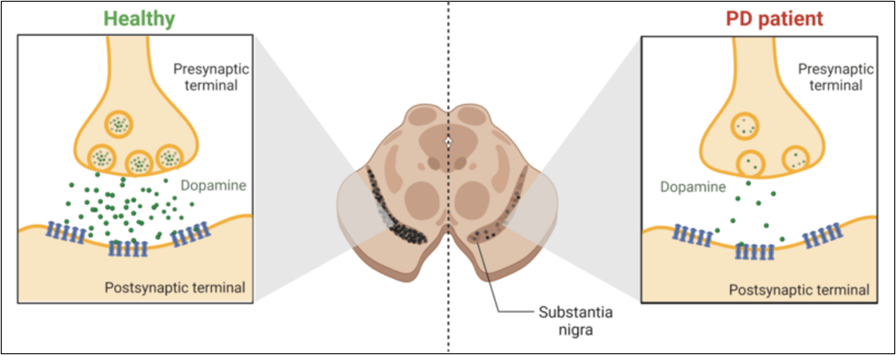
Programmed cell death, better known as apoptosis, is crucial in eliminating abnormal or unwanted cells in the body.[2] In recent years, the complex pathways regulating apoptosis have been well-studied as potential therapy targets.[2,10] Parkinson’s disease (PD) is no exception since the leading cause of this disorder is the death of dopamine-producing neurons in the brain (see Fig.1).[1-4] PD is a debilitating neurodegenerative disorder that steals a person’s ability to control movement.[8] Currently, levodopa (L-Dopa) is the most effective treatment for PD, helping in the improvement of the symptoms but not in the progression of the disease.[3] In addition, the use of L-Dopa led to adverse reactions after long-term administration.[3,7] Several studies propose using microRNAs (miRNAs) to inhibit the apoptosis of the dopamine-producing neurons found in PD.[6-9] miRNAs are tiny molecules capable of regulating gene expression (molecular switches) in the most critical processes for cell survival, like proliferation, cell differentiation, and apoptosis when required.[5-8]

Figure 1. The comparison between the normal functioning of dopaminergic neurons in healthy individuals vs in PD patients. 

For this reason, understanding the role of miRNAs in PD could be vital to developing new treatments to decrease the progression of the disorder. The apoptosis process is significant for eliminating unwanted cells.[2] Therefore, using miRNAs as a therapy has the potential to inhibit unwanted cell death and induce apoptosis in abnormal cells as well.[3-6] Understanding this interplay between miRNAs and apoptosis could lead to new treatment strategies for PD.[7] Several investigations found that miRNAs have neuroprotection abilities, safeguarding neurons from apoptotic cell death.[5] Alternatively, miRNAs promote apoptosis, thus eliminating damaged or dysfunctional neurons, which is crucial to clear cellular debris from microenvironments.[5]

Some benefits of using miRNAs as a therapy for PD are the following: (1) miRNAs can target specific genes involved directly in the apoptotic pathway (specificity). (2) miRNAs are molecular switches, activating or inhibiting the apoptosis pathway. This adaptive characteristic is very convenient for this type of therapy since PD requires, in some cases, the inhibition of neuronal death (dopamine-producing neurons) and the activation of apoptosis in unwanted cells in the brain. (3) miRNAs can cross the blood-brain barrier and enter into cells, which is a challenge in developing neurodegenerative diseases. The apoptosis pathway accelerates the progression of PD by the direct dopamine depletion caused by the death of dopamine-producing neurons.[4-6] As the disease progresses, more and more neurons are lost, leading to worsening symptoms and disability.

Stem Cells Show Promise for Alzheimer’s and Parkinson’s Disease.

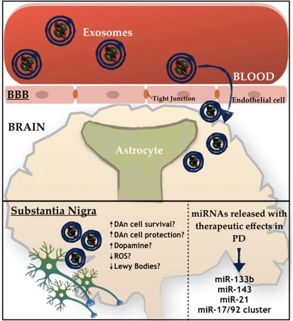
Mesenchymal stem cell-derived exosomes as a promising therapy for Parkinson’s and Alzheimer’s Disease.

Recently, investigators suggested using Mesenchymal stem cells (MSCs)–derived exosomes as a therapy for different conditions, including Parkinson’s Disease (PD).[1,2,5] MSCs can be found in various body parts and specialize in different cell types depending on the body’s needs.[1] These cells produce extracellular vesicles called exosomes that have been studied as an alternative medicinal agent because of their stability and biological prospect in terms of the substances they carry, like signaling molecules, cytokines, enzymes, and micro-RNA (miRNA).[1,2,4] All these components are essential in maintaining cellular homeostasis, while the miRNA is more involved in regulating gene expression. [1,2,4] Many studies with MSCs have demonstrated several benefits in other neuropathological conditions. [1] One of the insights is that the MSCs have been pointed to activate different neuro-regeneration processes, opening a door for many possible ways to serve as promising therapies for future clinical trials. [1,3] Two targets for developing new treatments using MSCs are PD and Alzheimer’s disease (AD). [2,3,4,7] PD is characterized by the deterioration of dopaminergic neurons and the insufficiency of dopamine production. [3,6,9] Generally, the decrease of dopaminergic neurons is related to the accumulation of Lewy bodies (protein aggregates of α-synuclein) inside the neurons, which affects the normal functioning of those cells.[9] Interestingly, MSCs-derived exosome seems to be able to decrease one of the leading causes of PD, neuroinflammation.[2,5,10] On the other hand, AD is described as a brain illness that presents as neurological hallmarks the formation of amyloid plaques (Aβ) and neurofibrillary tangles causing synaptic loss.[4,7,12]

Cerebrolysin Research

Cerebrolysin Constituents

Nerve Growth Factor: An important protein for development and maintenance of neurons, specifically, neurons involved in pain, temperature, and touch sensation. Nerve growth factor functions by binding to either the NTRK1 receptor or the p75NTR receptor which are both found on the surface of sensory neurons.Brain-Derived Neurotrophic Factor (BDNF): Protein found in the brain and spinal cord that is responsible for growth, maturation, and maintenance of neurons. In the brain, BDNF is active in the synapse and is important in regulating synaptic plasticity.Ciliary Neurotrophic Factor (CNTF): A brain derived protein that promotes neuron survival and axonal outgrowth during neuronal development.Enkephalins: Functions as a ligand for the kappa-type opioid receptor. Competes with and mimics effects of opiate drugs and therefore plays a role in pain perception and response to Stress.Orexin: Functions as a ligand which binds to orphan G-protein coupled receptors that play a role in regulation of sleep and arousal as well as feeding behavior, metabolism and Homeostasis.P21: The pentamer Ac-DGGL AG-NH2 is a peptide fragment of Cerebrolysin that has been found to play a role in neurogenesis and neuronal plasticity.

Cerebrolysin has been shown to help with (according to research):

• Alzheimer’s
• Vascular dementia
• Stroke recovery
• Traumatic brain injury (TBI) recovery
• Peripheral neuropathy
• Multiple scoliosis (MS)
• Parkinson’s

The Role of Cell-Permeable Peptides and The JNK Family in Preventing Neuronal Degeneration

Figure 1:     Neuroprotective features of CPPs in combination with other molecules or peptides for treating PD and AD.
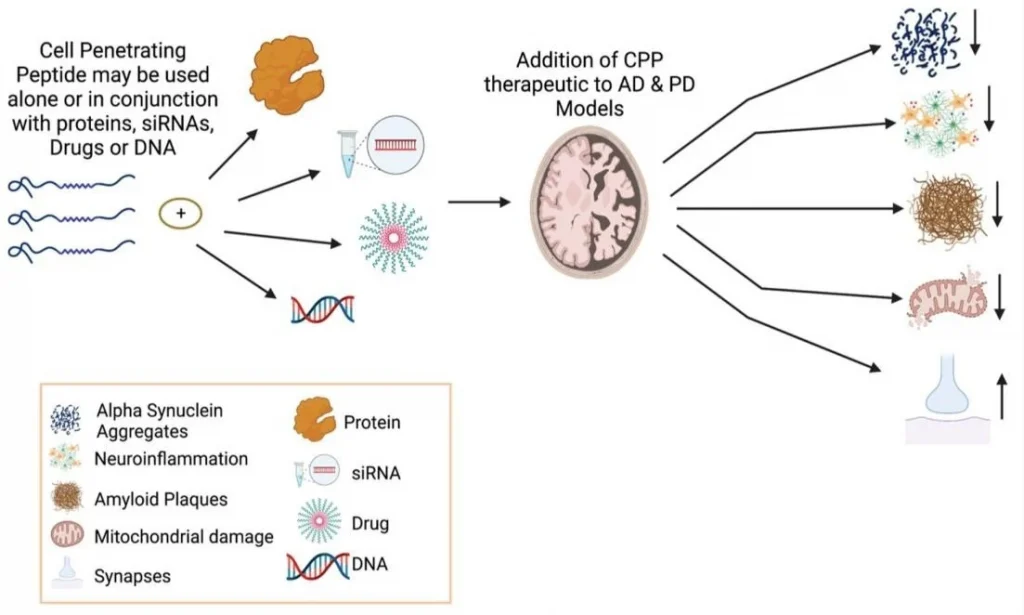
JNK family participates in the apoptosis pathway, also known as programmed cell death.[4] The apoptosis mechanism is used to eliminate cells that are damaged in anyway. This mechanism is suitable for healthy people because it helps avoid developing cancer and other disorders.[1] On the other hand, apoptosis is detrimental in neurodegenerative diseases that share the abnormal accumulation of misfolded proteins, contributing to dementia, cognitive loss, memory loss, behavioral problems, and sleep problems, among others, through neuronal cell loss.[3,5,7] In addition, the JNK family also participates in pathways related to regulation, plasticity, development of the Central Nervous System (CNS), inflammation, and autophagy.[9] The well-functioning of all these processes is vital. Recent studies suggested that an imbalance of the JNK family members can accelerate the progression of both AD and PD pathologies. One of the hallmarks of AD is the aggregation of amyloid beta (Aβ) in the extracellular area.[7] The overexpression of Aβ triggers the activation of JNK-3, which also participates in the formation of Aβ42, a toxic species that affects the neuron’s cell function when it accumulates.[7] It is unclear whether the neurodegenerative disorder or the JNK imbalance comes first. However, they both have a direct relationship where PD and AD patients show high levels of JNK in the brain (postmortem), and irregular levels of the JNK family accelerate the progression of both disorders (see Fig. 2). [5,9] JNK imbalance could also lead to increased inflammation responses, low plasticity, and a flaw in autophagy performance, among other related adverse effects. An increase in apoptosis in PD and AD patients causes a decrease in neurons in the brain, affecting cognitive, behavioral, and memory performance.[8] JNK also decreases the α-syn accumulation in PD models, contributing to neuroprotection.[10] JNK pathway influence positively many vital processes of the body unless an imbalance occurs. [2,8] JNK has been the target for treating several diseases like cancer, strokes, PD, AD, Huntington’s disease, and other neurodegenerative disorders, showing promising results. More investigations need to be done to comprehend better how the correct levels of JNK can help combat important hallmarks of degenerative illnesses to eventually prevent or revert the progression of related diseases like PD and AD. In general, the benefits of inhibiting JNKs for treating neurodegenerative disorders are:

­Improving autophagy for the elimination of misfolded proteins (Aβ (AD), tau protein (AD), and α-syn (PD)

  • ­Increasing cell proliferation.
  • Decreasing programmed cell death.
  • Reducing brain damage.
  • Decreasing cognitive decline.
  • Increasing gene expression.
  • Decreasing neuroinflammation responses
  • Improving memory
  • Providing neuroprotection
  • Improving synaptic connections

Welcome back!

or
Menu
Explore

Search Peptides

Welcome back!

or
0