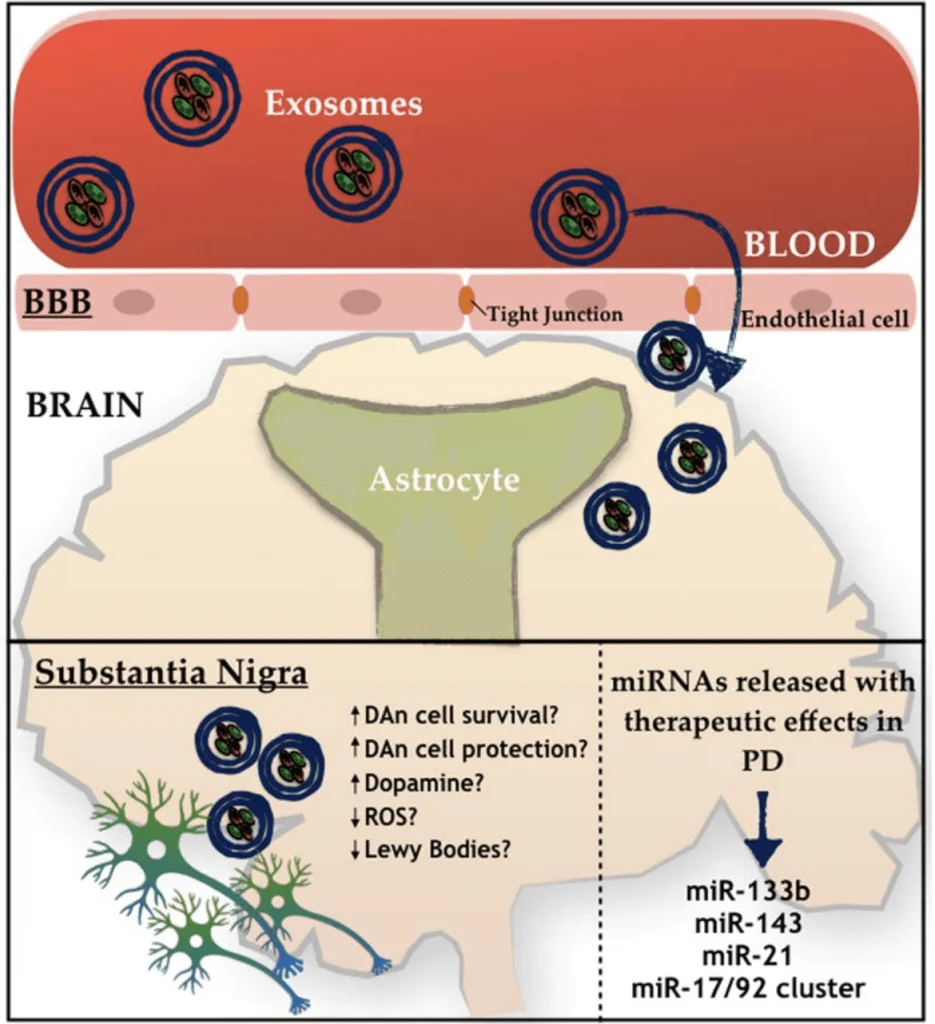
FREE SHIPPING ON ORDERS OVER $100

Research Categories
Research Categories

Stem Cell Research

ALL ARTICLES AND PRODUCT INFORMATION PROVIDED ON THIS WEBSITE ARE FOR INFORMATIONAL AND EDUCATIONAL PURPOSES ONLY. The products offered on this website are furnished for in-vitro studies only. In-vitro studies (Latin: in glass) are performed outside of the body. These products are not medicines or drugs and have not been approved by the FDA to prevent, treat or cure any medical condition, ailment or disease. Bodily introduction of any kind into humans or animals is strictly forbidden by law.

Stem Cells Show Promise for Alzheimer’s and Parkinson’s Disease.

Mesenchymal stem cell-derived exosomes as a promising therapy for Parkinson’s and Alzheimer’s Disease.

Recently, investigators suggested using Mesenchymal stem cells (MSCs)–derived exosomes as a therapy for different conditions, including Parkinson’s Disease (PD).[1,2,5] MSCs can be found in various body parts and specialize in different cell types depending on the body’s needs.[1] These cells produce extracellular vesicles called exosomes that have been studied as an alternative medicinal agent because of their stability and biological prospect in terms of the substances they carry, like signaling molecules, cytokines, enzymes, and micro-RNA (miRNA).[1,2,4] All these components are essential in maintaining cellular homeostasis, while the miRNA is more involved in regulating gene expression. [1,2,4] Many studies with MSCs have demonstrated several benefits in other neuropathological conditions. [1] One of the insights is that the MSCs have been pointed to activate different neuro-regeneration processes, opening a door for many possible ways to serve as promising therapies for future clinical trials. [1,3] Two targets for developing new treatments using MSCs are PD and Alzheimer’s disease (AD). [2,3,4,7] PD is characterized by the deterioration of dopaminergic neurons and the insufficiency of dopamine production. [3,6,9] Generally, the decrease of dopaminergic neurons is related to the accumulation of Lewy bodies (protein aggregates of α-synuclein) inside the neurons, which affects the normal functioning of those cells.[9] Interestingly, MSCs-derived exosome seems to be able to decrease one of the leading causes of PD, neuroinflammation.[2,5,10] On the other hand, AD is described as a brain illness that presents as neurological hallmarks the formation of amyloid plaques (Aβ) and neurofibrillary tangles causing synaptic loss.[4,7,12]

Peptide P21 slows the progression of neurodegeneration and Alzheimer’s by removing Tau protein build-up and reducing the production of Beta Amyloid plaques.

In Alzheimer’s disease, degeneration of brain synapses happens before Beta Amyloid plaques and Tau protein aggregates are produced.

“Both in AD and in its animal models the loss of neuronal plasticity is known to precede any overt formation of Aβ plaques and hyperphosphorylated (p) tau neurofibrillary tangles.” (1)

“Alzheimer’s disease responds to neurodegeneration by initiating neurogenesis in the dentate gyrus which, however, due to a lack of the proper neurotrophic support, is not sustained and the newborn neurons do not mature into functional cells.” (1)


“Alzheimer’s disease is characterized by neurodegeneration associated with loss of neuronal plasticity and in the dentate gyrus, proliferation of newborn cells which do not mature into functional neurons…” (1)


“Two major therapeutic approaches to Alzheimer’s disease and related conditions.

While one therapeutic approach to Alzheimer’s disease is the inhibition of neurodegeneration that is associated with neurofibrillary and Aβ pathologies, another approach is to stimulate the regeneration of the brain by enhancing neuronal plasticity and neurogenesis that culminates into formation of mature functional neurons.” (1)

Proper neurogenesis (birth of new neurons) by peptide P021 was shown to remove Tau protein aggregates and reduce the production of new Beta Amyloid plaques.
“Moreover, the P021 treatment markedly reduced tau pathology and attenuated the generation but not the clearance of Aβ in 3xTg-AD mice.” (2)

“Cognitive performance was studied by assessing episodic memory with Novel Object Recognition task at 16-17-months post-treatment. We found that P021 treatment initiated during the synaptic compensation period can prevent neurodegeneration, Aβ and tau pathologies, rescue episodic memory impairment, and markedly reduce mortality rate. These findings for the first time show effective prevention of AD changes with a neurotrophic compound that targets neurogenesis and synaptic plasticity, suggesting that improving the health of the neuronal network can prevent AD.” (3)

“The AD brain responds to neurodegeneration by stimulating neurogenesis, however, because of the lack of a proper neurotrophic microenvironment of the hippocampus, this effort of the AD brain to replace lost neurons with new neurons is unsuccessful and culminates in failure of neuronal survival, maturation, and integration. As the disease progresses, the neurogenic failure becomes severe, and contributes significantly to cognitive decline.” (4)

Posted in:

How does Thymosin Beta 4 (TB-500) enhance hair follicle growth?

Thymosin Beta 4 Increases Hair Growth by Activating Hair Follicle Stem Cells.

“Thymosin β4, a ubiquitous 4.9‐kDa polypeptide originally isolated from bovine thymus, is a potent mediator of cell migration and differentiation. It was identified as a gene up‐ regulated four‐ to sixfold during early endothelial cell tube formation and found to promote angiogenesis. It is present in wound fluid, and when added topically or given systemically, it promotes angiogenesis and wound healing. Thymosin β4 elicits cell migration through a specific interaction with actin. In angiogenesis and in wound healing, thymosin β4 acts by accelerating the migration of endothelial cells and keratinocytes and increasing the production of extracellular matrix‐degrading enzymes.”
“Thymosin β4 promotes hair growth in normal rats and mice. A specific subset of follicular keratinocytes in the mouse skin, which originates at the bulge region, expresses thymosin β4. The temporal and spatial distribution of these keratinocytes parallel the pattern reported for the stem cells and their daughter TA cells at the different stages of the hair cycle (910). We isolated clonogenic keratinocytes from the bulge compartment of the rat vibrissa follicle, further characterized them as an immediate progeny of the stem cells, and found that these cells express high levels of thymosin β4 when cultured in vitro. We show that thymosin β4 promotes hair clonogenic keratinocyte cell migration, as well as secretion of the extracellular matrix‐degrading enzyme matrix metalloproteinase 2 (MMP‐2).””Thus, thymosin β4 accelerates hair growth, in part, due to its effect on critical events in the active phase of the hair follicle cycle, including promoting the migration of stem cells and their immediate progeny to the base of the follicle, differentiation, and extracellular matrix remodeling.”
“Taken together, our results suggest that in addition to its known angiogenic and wound healing effects, thymosin β4 is a naturally occurring modulator of hair growth that acts by stimulation of stem cell migration, protease production, and differentiation.”

“While studying wound healing in rat skin, we unexpectedly observed visually and at the histological level increased hair growth at the wound margins 7 days after topical treatment with thymosin β4 (unpublished observation). In this study, we have shaved the skin of healthy rats and applied thymosin β4 topically on one side of the shaved area and the control vehicle on the opposing lateral side of the same animal. After 7 days of treatment, we observed an increased number of anagen‐phase hair follicles in the skin areas treated with thymosin β4 (Fig. 1a and d). The number of anagen follicles was approximately twofold greater than in rats treated with vehicle alone. The increased number of hairs in anagen phase was retained with continued tri‐ weekly treatment over 30 days. Within 14 days of treatment cessation, the number of active hair follicles decreased to control levels. We next tested whether thymosin β4 would promote hair growth in 8‐wk‐old C57BL6 wild‐type mice. Animals used in this experiment have all of their hair follicles in the telogen stage as judged by their pink skin color. The mice were shaved and thymosin β4 was applied topically on the shaved area as described in Methods. Control animals were treated with vehicle alone. As shown in Fig. 1c and ƒ, thymosin β4‐treated (but not control) animals displayed quick hair regrowth. Histological examination confirmed the thymosin β4‐induced activation of the hair follicles (Fig. 1b and e).”

Peptides and Stem Cell Research

Thymosin Beta-4

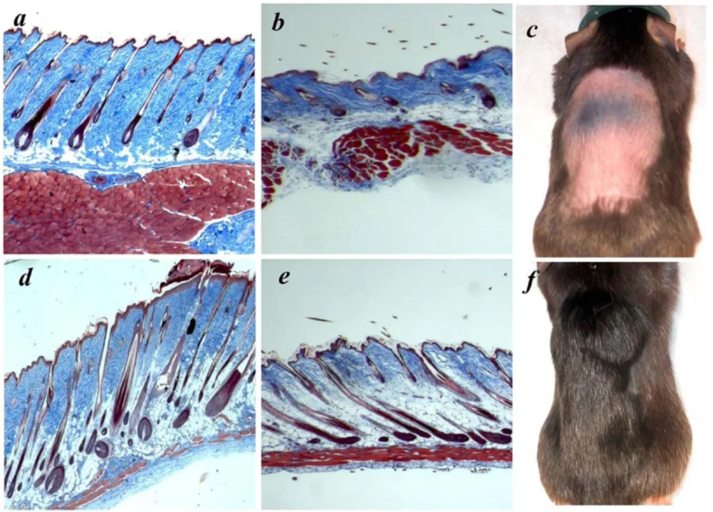
Thymosin Beta 4, is a member of a highly conserved family of actin monomer-sequestering proteins. Thymosin β-4 is a 43 amino acid sequence encoded by gene TMSBX4 present in all human cells. It is naturally found in higher concentrations in tissue damaged areas and has been frequently used in sports doping for the past 20 years for its ability to decrease injury times and reduce delayed onset muscle soreness. TB-4 has been used for soft tissue repair (this includes tendon, ligament, and muscle), sports and athletic injuries, pressure or venous stasis ulcers, immune response modulation, brain issues related to autoimmunity, and spinal cord injuries. In addition to its role as a major actin-sequestering molecule, Thymosin Beta 4 plays a role in tissue repair. Tβ4 has been found to play an important role in protection, regeneration and remodeling of injured or damaged tissues. The gene for Tβ4 has also been found to be one of the first to be upregulated after injuries. Thymosin Beta 4 is most often prescribed for acute injury, surgical repair and for senior athletes. It has most recently been shown to help regrow hair in addition to PRP and has several effects on stem cell activation.

Advances in the basic and clinical applications of thymosin beta 4:

Based on its multifunctional activities during tissue regeneration in various animal studies in this report, Tb4 has the potential for new clinical applications such kidney and liver disease, as well as repair of spinal cord, bone and ligament damage.

The effect on stem cells:

Thymosin beta-4 promotes mesenchymal stem cell proliferation via an interleukin-8-dependent mechanism:

This study shows that Tβ4 promotes the expansion of human ASCs via an IL-8-dependent mechanism that involves the ERK and NF-κB pathways. Therefore, Tβ4 could be used as a tool for MSC expansion in cell therapeutics.

Thymosin Beta-4 Directs Cell Fate Determination of Human Mesenchymal Stem Cells through Biophysical Effects:

Tb4 initiated cell fate determination of MSCs through biophysical effects exerted by cytoskeleton reorganization and altered cell-cell adhesion rather than direct regulation of lineage-determining transcription factors.

Neuroprotective and neurorestorative effects of Thymosin beta 4 treatment following experimental traumatic brain injury:

Delayed (24 hours post injury) Tβ4 treatment promotes neurogenesis after TBI in rats.

Thymosin beta 4 up-regulates miR-200a expression and induces differentiation and survival of rat brain progenitor cells:

The miR-200 not only induces terminal differentiation of NPCs in the OB, but also induces neuronal differentiation in neuronal stem cells and in a PC-12 cell line.

PEG-MGF

MGF is a split variant of IGF-1 but its sequence differs from the systemic IGF-1 produced by the liver. MGF initiates hypertrophy and repair of local muscle damage. MGF is expressed by mechanically overloaded muscle and is involved in tissue repair and adaptation. It is expressed as a pulse following muscle damage and is involved in the activation of muscle satellite (stem) cells. These donate nuclei to the muscle fibers that are required for repair and for the hypertrophy process, which may have similar regulatory mechanisms. MGF is essential for repair and therefore growth of new cells, similar to IGF-1.

The effect on stem cells:

Mechano growth factor E peptide regulates migration and differentiation of bone marrow mesenchymal stem cells:

Both wound-healing and transwell assays indicated that MGF E peptide could significantly enhance rBMSCs migration ability.

Welcome back!

or
Menu
Explore

Search Peptides

Welcome back!

or
0