FREE SHIPPING ON ORDERS OVER $100

Research Categories
Research Categories

Telomere Research

ALL ARTICLES AND PRODUCT INFORMATION PROVIDED ON THIS WEBSITE ARE FOR INFORMATIONAL AND EDUCATIONAL PURPOSES ONLY. The products offered on this website are furnished for in-vitro studies only. In-vitro studies (Latin: in glass) are performed outside of the body. These products are not medicines or drugs and have not been approved by the FDA to prevent, treat or cure any medical condition, ailment or disease. Bodily introduction of any kind into humans or animals is strictly forbidden by law.

How does Epitalon enhance sleep while protecting DNA and Telomeres?

Epithalon Peptide Induces Telomerase Activity and Telomere Elongation in Human Somatic Cells and Overcomes the Hayflick Limit

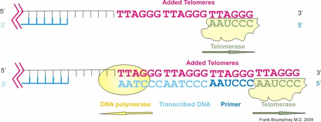
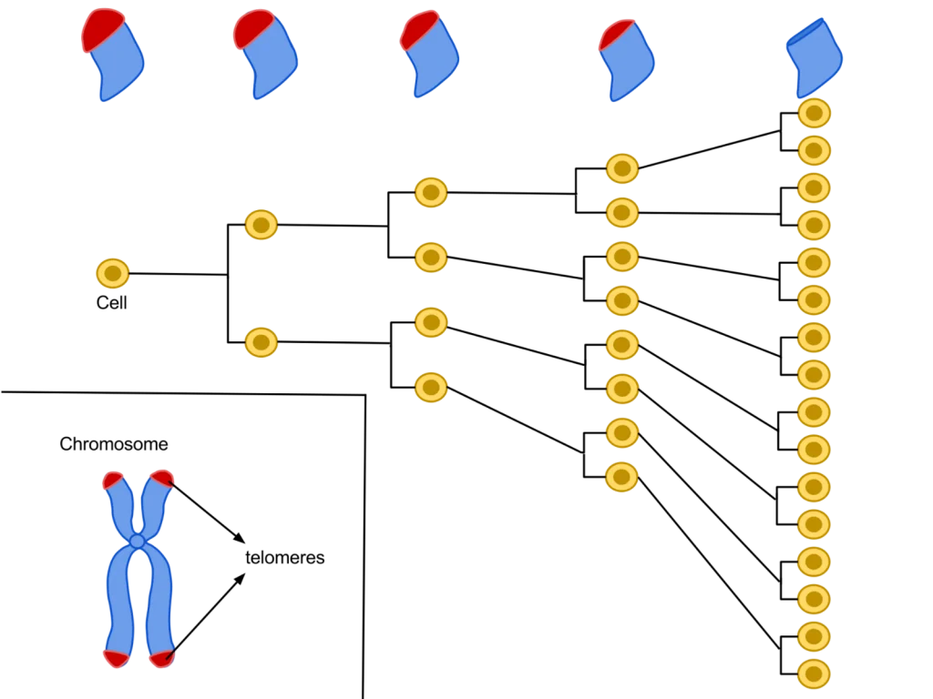
Each cell contains DNA as an instruction manual for how to divide and grow. The DNA inside of each cell is shielded by proteins called telomeres. During cellular division, a new cell must take some telomeres from its originating cell to shield the DNA of the new cell. The telomeres shorten after every cell division because the new cell can only take a portion of the telomeres from the previous cell, else the previous cell’s DNA will become completely unprotected.Once there are no left over telomeres to take, the cell stops dividing. This happens after a single cell divides and grows about 64 other cells, which is known as the Hayflick limit. This limit exists because cells without shield material are more vulnerable DNA damage. If the DNA of a cell becomes damaged, the cell will follow broken instructions. If the instructions within the DNA of the cell are damaged, then the cell may not be able to eliminate itself through the process of apoptosis like it is supposed to.”The telomere length is increased by approximately 33% in epitalon treated cells [by increasing the telomerase enzyme that strengthens telomeres].” (10)“Telomerase is a reverse transcriptase that has two distinct functions, to replicate pre-existing chromosome ends (telomeres) and to heal broken chromosomes by de novo addition of telomeric sequences directly on to non-telomeric DNA.” (11)

“Addition of Epithalon to aging cells in culture induced elongation of telomeres to the size comparable to their length during early passages. Peptide-treated cells with elongated telomeres made 10 extra divisions (44 passages) in comparison with the control and continued dividing. Hence, Epithalon prolonged the vital cycle of normal human cells due to overcoming the Hayflick limit.” (12)

Cellular Senescence and Chronic Pain

Senescence Induced Inflammation (SASP) May Promote Cell and Telomere Damage, Leading to Allodynia.

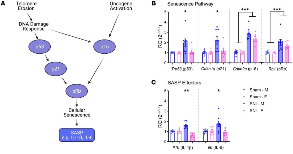
“Expression of senescence pathway and SASP effector genes in the spinal cords of mice of both sexes 6 months after sham or SNI surgery. “

(SASP is inflammation secreted by senescent cells.)

“Peripheral nerve injury produces cellular senescence in the spinal cord of mice at time points long after injury. Reduced TL can result in a persistent DNA damage response leading to cellular senescence — a state of cell cycle arrest/withdrawal, deregulated cellular metabolism, and macromolecular damage — and senescent cells in turn release a diverse set of cytokines, growth factors, proteases, and extracellular matrix components, together known as the senescence-associated secretory phenotype (SASP) or senescent-messaging secretome. Many of these SASP-related compounds are proinflammatory, and well known to produce or facilitate pain, especially when released in the spinal cord. We gave new cohorts of young male and female mice SNI or sham surgeries and harvested lumbar spinal cord tissues from these animals 12–14 months later, or 2 months later.” (1)

Posted in:

How Do Telomeres Impact Aging and Autophagy?

Loss of Telomere Leads to p53 and Autophagy Induced Cell Death.

“autophagy-deficient cells … continued to proliferate and bypassed crisis” (2)
“In summary, cells in telomere crisis undergo cell death through autophagy, which is triggered by chromosome breakage and transduced by the cGAS–STING pathway. As cell death in crisis represents the final barrier against neoplastic transformation, a cancer therapy that involves inhibition of autophagy could be counterproductive… Moreover, cells lacking either cGAS or STING proliferated beyond crisis. “”Autophagy mediates the turnover of cytoplasmic macromolecules to support cellular homeostasis. Autophagy generally blocks apoptosis, but in specific circumstances it can lead to cell death through excessive degradation of cell constituents. The authors studied telomere crisis using human fibroblasts and epithelial cells, in which the RB and/or p53 pathways were suppressed; these cells bypassed senescence and entered replicative stress, exhibiting telomere attrition, chromosome fusions and cell death.””Telomere deprotection through TRF2 depletion was sufficient to activate autophagy independently of replicative crisis, and genetic suppression of telomere fusions in TRF2-depleted cells reduced the accumulation of cytosolic DNA and attenuated autophagy, suggesting that fusion-dependent cytosolic DNA is required for the telomeric autophagy response. ” (2)

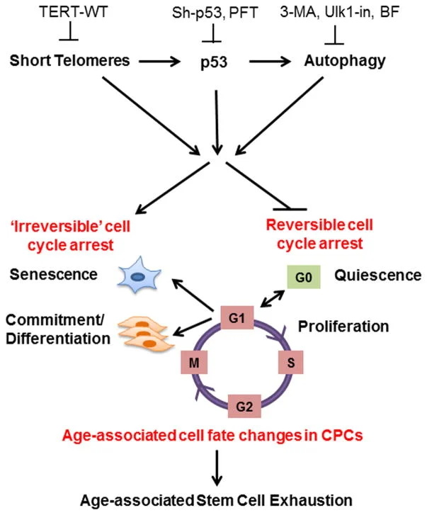
“The cell fate of CPCs changes with age and is characterized by a switch away from proliferation and quiescence (reversible form of cell cycle arrest) towards senescence and increased basal commitment (‘irreversible’ forms of cell cycle arrest) accounting for age-associated stem cell exhaustion. Mechanistically, short telomeres activate p53 that induces autophagy and at least partially contributes to the age-associated change in cell fate. Blunting telomere shortening via overexpression of TERT-WT, silencing p53 , or treating with pharmacological inhibitors of p53 (PFT) and autophagy (3-MA, Ulk1-In, BF) selectively attenuate senescence and basal commitment and reverse cell fate of aged CPCs.” (3)

Circadian Rhythm Controls Telomeres and Telomerase Activity.
“Circadian clocks are fundamental machinery in organisms ranging from archaea to humans. Disruption of the circadian system is associated with premature aging in mice, but the molecular basis underlying this phenomenon is still unclear. In this study, we found that telomerase activity exhibits endogenous circadian rhythmicity in humans and mice. Human and mouse TERT mRNA expression oscillates with circadian rhythms and are under the control of CLOCK–BMAL1 heterodimers.

CLOCK deficiency in mice causes loss of rhythmic telomerase activities, TERT mRNA oscillation, and shortened telomere length. Physicians with regular work schedules have circadian oscillation of telomerase activity while emergency physicians working in shifts lose the circadian rhythms of telomerase activity. These findings identify the circadian rhythm as a mechanism underlying telomere and telomerase activity control that serve as interconnections between circadian systems and aging.” (4)

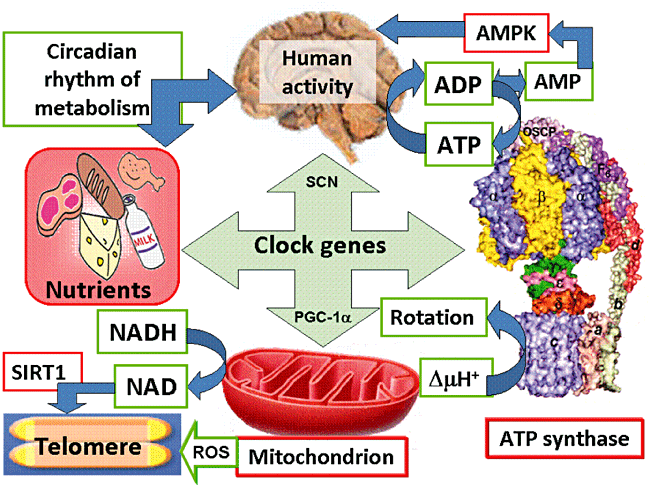
“Human activity is driven by NADH and ATP produced from nutrients, and the resulting NAD and AMP play a predominant role in energy regulation. Caloric restriction increases both AMP and NAD and is known to extend the healthspan (healthy lifespan) of animals. Silent information regulator T1 (SIRT1), the NAD-dependent deacetylase, attenuates telomere shortening, while peroxisome proliferator-activated receptor γ coactivator 1α (PGC-1α), a master modulator of gene expression, is phosphorylated by AMP kinase and deacetylated by SIRT1. Thus, PGC-1α is a key component of the circadian oscillator that integrates the mammalian clock and energy metabolism.

Reactive oxygen species produced in clock mutants result in telomere shortening. The circadian rhythms produced by clock genes and lifestyle factors are ultimately controlled by the human brain and drive homeostatic and hedonic feeding and daily activity. ” (9)

  • Telomerase and TERT mRNA expressions exhibit endogenous circadian rhythm.
  • Human and mouse TERT mRNA expression are under the control of CLOCK–BMAL1 heterodimers.
  • CLOCK deficient mice have shortened telomere length and abnormal oscillations of telomerase activity and TERT mRNA.
  • Emergency physicians working in shifts lose the circadian rhythms of telomerase activity.

What is Epithalon?

Epithalon is currently being studied and researched by Scientists and Doctors specializing in the field of anti-aging care and medicine. EPITHALON (Epitalon) is one of the most important breakthroughs in the study of anti-aging.

The Epithalon (Epitalon) tetrapeptide has been discovered by researchers in Russia. This was seen to reactivate the production of cell telomerase thus slowing down the aging process and rejuvenating the entire body. The development of molecular biology required bio-chemical studies that were nothing short of profound. Scientific work by Gobind Khorana and Marshall Nirenberg for many years resulted in defining codons or nucleotides and triplets and the genetic code of each of the 20 amino acids. This resulted in a Nobel Prize award in 1968 with Robert Holley. Nucleic acid investigations and identification of DNA and RNA base sequences were also conducted by the 1980 Nobel Prize winner for Chemistry Frederick Sanger along with Walter Gilbert and Paul Berg. These studies revealed the cause of aging.

Epithalon Fights Aging by Activating Telomerase

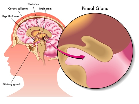
Epithalon, also known as Epitalon is a synthetic peptide analog of epithalamin, a protein found in the pineal gland of mammals and of interest for its anti-aging properties. Past research studies have demonstrated that epithalamin can increase maximum life span in animals, decrease levels of free radicals, and alter catalase activity to prevent tissue damage [1]. Epithalamin has been shown to decrease mortality by 52% in fruit flies, by 52% in normal rats, and by 27% in mice prone to certain types of cancer and cardiovascular disease [2].

It has been shown, through extensive animal research, to be a potent regulator of cell metabolism, including growth and cell division. In particular, epithalon is able to extend cell survival in vitro. At least part of the reason that epithalon can extend cell survival comes down to its action on telomeres.

Epithalon has similar effects to epithalamin in mice and rats. It has also shown promise as an anti-cancer agent, reducing spontaneous mammary tumors in mice prone to them and reducing incidence of intestinal tumors in rodents. How does it achieve these effects?

Welcome back!

or
Menu
Explore

Search Peptides

Welcome back!

or
0