FREE SHIPPING ON ORDERS OVER $100

Research Categories
[list_research_categories]
Research Categories

Research

ALL ARTICLES AND PRODUCT INFORMATION PROVIDED ON THIS WEBSITE ARE FOR INFORMATIONAL AND EDUCATIONAL PURPOSES ONLY. The products offered on this website are furnished for in-vitro studies only. In-vitro studies (Latin: in glass) are performed outside of the body. These products are not medicines or drugs and have not been approved by the FDA to prevent, treat or cure any medical condition, ailment or disease. Bodily introduction of any kind into humans or animals is strictly forbidden by law.

miRNA Researched for Parkinson’s Disease: Neuroprotection Blocks Neuron Death

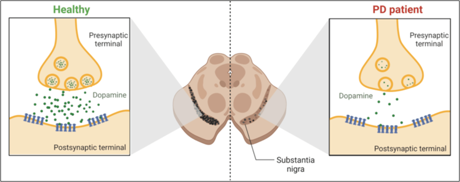
Programmed cell death, better known as apoptosis, is crucial in eliminating abnormal or unwanted cells in the body.[2] In recent years, the complex pathways regulating apoptosis have been well-studied as potential therapy targets.[2,10] Parkinson’s disease (PD) is no exception since the leading cause of this disorder is the death of dopamine-producing neurons in the brain (see Fig.1).[1-4] PD is a debilitating neurodegenerative disorder that steals a person’s ability to control movement.[8] Currently, levodopa (L-Dopa) is the most effective treatment for PD, helping in the improvement of the symptoms but not in the progression of the disease.[3] In addition, the use of L-Dopa led to adverse reactions after long-term administration.[3,7] Several studies propose using microRNAs (miRNAs) to inhibit the apoptosis of the dopamine-producing neurons found in PD.[6-9] miRNAs are tiny molecules capable of regulating gene expression (molecular switches) in the most critical processes for cell survival, like proliferation, cell differentiation, and apoptosis when required.[5-8]

Figure 1. The comparison between the normal functioning of dopaminergic neurons in healthy individuals vs in PD patients. 

For this reason, understanding the role of miRNAs in PD could be vital to developing new treatments to decrease the progression of the disorder. The apoptosis process is significant for eliminating unwanted cells.[2] Therefore, using miRNAs as a therapy has the potential to inhibit unwanted cell death and induce apoptosis in abnormal cells as well.[3-6] Understanding this interplay between miRNAs and apoptosis could lead to new treatment strategies for PD.[7] Several investigations found that miRNAs have neuroprotection abilities, safeguarding neurons from apoptotic cell death.[5] Alternatively, miRNAs promote apoptosis, thus eliminating damaged or dysfunctional neurons, which is crucial to clear cellular debris from microenvironments.[5]

Some benefits of using miRNAs as a therapy for PD are the following: (1) miRNAs can target specific genes involved directly in the apoptotic pathway (specificity). (2) miRNAs are molecular switches, activating or inhibiting the apoptosis pathway. This adaptive characteristic is very convenient for this type of therapy since PD requires, in some cases, the inhibition of neuronal death (dopamine-producing neurons) and the activation of apoptosis in unwanted cells in the brain. (3) miRNAs can cross the blood-brain barrier and enter into cells, which is a challenge in developing neurodegenerative diseases. The apoptosis pathway accelerates the progression of PD by the direct dopamine depletion caused by the death of dopamine-producing neurons.[4-6] As the disease progresses, more and more neurons are lost, leading to worsening symptoms and disability.

What is Epithalon and How Does it Protect Telomeres?

What is Epithalon?

Epithalon is a short, 4 amino acid chain peptide used to regulate the cell cycle through the upregulation of telomerase activity. It has been shown to have distinctive anti-aging and anti-tumor activity across many animal and human studies. Known as the synthetic version of the tetrapeptide epithalamin, which naturally occurs in the pineal gland in our body, Epithalon (also known as Epitalon or Epithalone) was first discovered in the late 1980’s by Prof. Vladimir Khavinson from The Sankt Petersburg University, Russia.

As the most prominent tasks of the pineal gland are to maintain different kind of processes in our body, such as to normalize the activity of anterior pituitary and to maintain the levels of calcium, gonadotropins, and melatonin, its activity is highly regulated by a series of feedback mechanisms. Epithalamin acts as an antioxidant and increases the resistance to stress and lowers the levels of corticosteroids. The life extension and anti-aging properties, amongst a variety of different clinical indications, of epithalon are incredible.

Scientific research has revealed that epithalon affects the following:

– Upregulate telomerase activity
– Normalize antioxidant indices
– Reduce peroxide lipid oxidation products
– Increase activity of glutathione peroxidase
– Improve melatonin and immunity (cellular and humoral)
– Improve insulin sensitivity
– Decrease LDL and VLDL
– Improve tissue repair
– Anti-tumor effects
– Decrease mortality and increases life expectancy


Epithalon and Skin Rejuvenation

Epithalon and Skin Rejuvenation

Skin rejuvenation is often associated with wrinkles and lines, but the truth runs deeper than wrinkles. Skin becomes more fragile and thus more prone to damage as it ages. Damage to the skin compromises its protective barrier function and can increase risk of infection. Research into ways to strengthen skin can not only make skin look younger, but can protect people from serious medical conditions. Thus far, most skin rejuvenation research has focused on collagen and other large skin proteins. New research, however, suggests that short peptide molecules, like epithalon, may hold more promise in preserving and even rejuvenating skin.

Epithalon Overview

Epithalon (a.k.a. epitalon), is a short (just four amino acids long) peptide that has been demonstrated to have anti-aging and anti-cancer properties in rodent studies. Because epithalon is so short, it can penetrate the cell membrane, without the aid of transporters, and make its way to the nucleus of cells. This is important because, once in the nucleus, epithalon can affect the regulation of genes, activating some and deactivating others to cause cell-wide changes1.

Previous research has indicated that epithalon can stimulate immune system function that has been lost due to natural aging. Investigation of the mechanism of this action uncovered the ability of the Ala-Glu-Asp-Gly peptide chain (Epithalon) to interact with the promoter region of the interferon gamma gene. By promoting the production of interferon gamma, a key immune regulator, epithalon is able to boost functioning in T-cells and thus overall immunity and well being1,2.

The idea that short peptides might be able to affect DNA-level processes has caused a boom in the investigation and research of epithalon and other short peptides in animal models. Those investigations hav

What is Thymosin Beta 4, How Does it Work?

What is Thymosin Beta-4?

The beta-thymosins (b-thymosins) comprise a family of structurally related, highly conserved amino acid sequences in species ranging from mammals to echinoderms. Of the 16 known family members, thymosin β4 (Tb4), thymosin β10 (Tb10), and thymosin β15 (Tb15) are found in man.

Thymosin beta-4 (TB4) is a 43 amino acid, 5kDa polypeptide that is an important mediator of cell proliferation, migration, and differentiation. TB4 is the most abundant member of the β- thymosin family in mammalian tissue and is regarded as the main G-actin sequestering peptide. It is found in all tissues and cell types except red blood cells. Thymosin beta-4 is angiogenic and can promote endothelial cell migration and adhesion, and angiogenesis. TB4 also accelerates wound healing and reduces inflammation and scarring when applied in dermal wound-healing assays.

Beta thymosins bind and sequester monomeric actin, thus preventing actin polymerization and formation of filamentous actin. Actin is a vital component of cell structure and movement. Actin is involved in many important non-muscle cellular processes, including cell locomotion, chemotaxis, phagocytosis, and cytokinesis. Of the thousands of proteins present in cells, actin makes up to 10% of the total proteins in a cell, representing a major role in the genetic makeup of the cell.

Animal studies of disease and repair when using thymosin beta-4, the major actin-sequestering molecule in mammalian cells, have provided a base for the ongoing multicenter clinical trials for wound healing, including dermal, corneal, and cardiac. TB4 has multiple biological activities, which include down-regulation of inflammatory chemokines and cytokines, and promotion of cell migration, blood vessel formation, cell survival, and stem cell maturation.

Thymosin beta-4 also inhibits inflammation, microbial growth, scar formation (by reducing the level of myofibroblasts), and apoptosis, and protects cells from cytotoxic damage, including glutamate neuronal toxicity. In addition, it binds to G-actin, blocks actin polymerization, and is released with factor X by platelets. These activities contribute to the multiple wound healing properties that have been reported in animal and human studies.

BPC-157 as Potential Support for Viral Infections

BPC 157 is a peptide that has demonstrated anti-inflammatory, cytoprotective, and endothelial-protective effects in different organ systems in different species. BPC 157 activated endothelial nitric oxide synthase (eNOS) is associated with nitric oxide (NO) release, tissue repair and angio-modulatory properties which can lead to improved vascular integrity and immune response, reduced proinflammatory profile, and reduced critical levels of the disease. As a result, discussion of its use as a potential prophylactic and complementary treatment is critical.

Figure 2: BPC 157 Molecule

Researchers hypothesize BPC 157 to be a promising future treatment for COVID-19 patients. Plausibly, BPC 157 may offer improved COVID-19 outcomes by mitigating cytokine derailment and subsequent multi-organ failure based on its anti-inflammatory, cytoprotective, and endothelium-protecting effects (e.g., through BPC 157-eNOS interactions). Furthermore, BPC 157 applications may obstruct viral replication, improve clinical and biochemical parameters, attenuate organ damage from the systemic alterations, provoked from SARS-CoV-2. Support for such a hypothesis is explained in further detail below.

How can Thymosin Beta4 (TB500) improve recovery, inflammation, neuropathies, fibrosis, telomerase and senescent cell removal?

Thymosin B4 Reduces Inflammation by Upregulating MicroRNA-146a and Promotes Myelin

“Tissue inflammation results from neurological injury, and regulation of the inflammatory response is vital for neurological recovery. The innate immune response system, which includes the Toll-like receptor (TLR) proinflammatory signaling pathway, regulates tissue injury… TB4-mediated oligodendrogenesis results from [up-regulating] miR-146a [causing the] suppression [of] the TLR proinflammatory pathway and modulation of the p38 MAPK pathway.” (8)“By targeting IRAK1 and TRAF6, miR-146 inhibits NF-κB activation. We therefore hypothesized that TB4 regulates the TLR proinflammatory signaling pathway by specifically regulating miR-146a to promote differentiation of OPCs [oligodendrocyte progenitor cells] to mature myelin basic protein (MBP)-expressing OLs [oligodendrocytes]… transfection with anti-miR-146a inhibitor nucleotides significantly inhibited the expression of MBP and phosphorylation of p38 MAPK.” (8)

How does Epitalon enhance sleep while protecting DNA and Telomeres?

Epithalon Peptide Induces Telomerase Activity and Telomere Elongation in Human Somatic Cells and Overcomes the Hayflick Limit

Each cell contains DNA as an instruction manual for how to divide and grow. The DNA inside of each cell is shielded by proteins called telomeres. During cellular division, a new cell must take some telomeres from its originating cell to shield the DNA of the new cell. The telomeres shorten after every cell division because the new cell can only take a portion of the telomeres from the previous cell, else the previous cell’s DNA will become completely unprotected.Once there are no left over telomeres to take, the cell stops dividing. This happens after a single cell divides and grows about 64 other cells, which is known as the Hayflick limit. This limit exists because cells without shield material are more vulnerable DNA damage. If the DNA of a cell becomes damaged, the cell will follow broken instructions. If the instructions within the DNA of the cell are damaged, then the cell may not be able to eliminate itself through the process of apoptosis like it is supposed to.”The telomere length is increased by approximately 33% in epitalon treated cells [by increasing the telomerase enzyme that strengthens telomeres].” (10)“Telomerase is a reverse transcriptase that has two distinct functions, to replicate pre-existing chromosome ends (telomeres) and to heal broken chromosomes by de novo addition of telomeric sequences directly on to non-telomeric DNA.” (11)

“Addition of Epithalon to aging cells in culture induced elongation of telomeres to the size comparable to their length during early passages. Peptide-treated cells with elongated telomeres made 10 extra divisions (44 passages) in comparison with the control and continued dividing. Hence, Epithalon prolonged the vital cycle of normal human cells due to overcoming the Hayflick limit.” (12)

BPC-157 vs TB-500

BPC-157 vs TB-500

BPC-157 and TB-500 are both potent healing peptides with vast amounts of research investigating their properties and potential uses. Both are also synthetic derivatives of naturally occurring proteins that have been modified to enhance their already abundant features. Both peptides have been shown to improve immune function, enhance healing throughout the body, and even thwart the ravages of age in some ways. Still, BPC-157 vs TB-500 is a valid comparison as these two peptides are not the same and do not share all of the same functions. Below is a look at why someone might choose BPC-157 over TB-500 or vice versa. 

BPC-157 vs TB-500: General Wound Healing

Both TB-500 and BPC-157 have been shown to accelerate wound healing and tissue repair. BPC-157, a derivative of body protection compound (BPC), has a dose-dependent effect on the growth and migration of fibroblasts, the cells responsible for extracellular matrix repair[1]. TB-500, a derivative of thymosin beta-4 (Tβ-4) has a similar effect that it produces by manipulating actin filaments. Actin is a protein that plays central roles in cell reproduction and migration. Research shows that TB-500 can increase the rate of fibroblast growth and migration as well as boost health and migration of cells of the immune system.

Hallmarks of Aging Part 1 of 4

Telomere Attrition

Telomere attrition is a hallmark of aging that refers to the shortening of telomeres, which are the protective caps on the ends of chromosomes. Telomeres play a critical role in maintaining the stability of the genome and protecting DNA from damage. With each cell division, telomeres become shorter, eventually leading to cellular senescence or cell death.

Telomere attrition is important because it is thought to contribute to the aging process and the development of age-related diseases. As telomeres become shorter, cells become more vulnerable to DNA damage, which can lead to cellular dysfunction and contribute to the development of diseases such as cancer. In addition, shortened telomeres have been linked to a range of age-related diseases, including cardiovascular disease, dementia, and diabetes.

The role of telomere attrition in aging is complex and not fully understood. However, it is thought that telomere shortening contributes to the decline in physiological function that characterizes aging. This may be due to the loss of key cellular processes, such as stem cell function and immune system function, that are necessary for maintaining tissue homeostasis

There are several strategies that have been proposed to address telomere attrition and promote healthy aging. One approach is to enhance telomerase activity, which is the enzyme responsible for maintaining telomere length. This has been shown to slow down telomere shortening and promote cellular longevity in some studies. Another potential strategy is to reduce exposure to factors that contribute to telomere shortening, such as oxidative stress and inflammation.

In addition, lifestyle factors have been shown to play a role in telomere length maintenance. For example, regular exercise has been associated with longer telomere length, while smoking and poor diet have been associated with shorter telomeres. Therefore, adopting healthy lifestyle habits may also help to promote healthy aging and protect against telomere attrition.

Telomere attrition is one of the 12 hallmarks of aging that can contribute to cellular dysfunction and the development of age-related diseases. While the mechanisms underlying telomere attrition and its role in aging are complex and not fully understood, strategies to address telomere attrition, such as enhancing telomerase activity and adopting healthy lifestyle habits, show promise as potential interventions for promoting healthy aging.


​Hallmarks of Aging Part 2 of 4

Hallmarks of Aging Part 2 of 4

As we age, our bodies undergo a complex series of changes that result in a decline in our overall health and an increased risk of age-related diseases. The aging process is multifaceted, and recent research has identified three key biological mechanisms that play a central role in this process. These mechanisms are Cellular Senescence, Mitochondrial Dysfunction, and Deregulated Nutrient Sensing.Cellular Senescence is a process in which cells become irreversibly arrested in a state of growth arrest, preventing them from dividing and contributing to tissue repair and regeneration. While cellular senescence can be a beneficial response to stress or damage in some cases, its chronic activation can lead to the accumulation of senescent cells in our tissues, which can contribute to inflammation and other harmful effects.Mitochondrial Dysfunction refers to the decline in the functioning of our mitochondria, which are the energy-producing organelles in our cells. As we age, the efficiency of our mitochondria decreases, leading to a reduction in energy production and an increase in the production of harmful byproducts known as reactive oxygen species.Deregulated Nutrient Sensing refers to the dysregulation of various signaling pathways that control our metabolism and nutrient uptake. This dysregulation can lead to the accumulation of harmful byproducts and the development of age-related diseases such as diabetes and cardiovascular disease.Understanding these three antagonistic hallmarks of aging is crucial for developing interventions and therapies that can improve health span and extend lifespan. Remember, “The antagonistic hallmarks of aging are hallmarks that can have beneficial or deleterious effects on the cell, depending on the level of intensity. When regulated properly, these hallmarks are beneficial or protective, but can be deleterious when levels are too high, or unregulated.” By targeting these mechanisms, researchers hope to develop strategies that can slow or even reverse the aging process, paving the way for healthier and more productive lives in old age.

Cellular Senescence

Cellular senescence is a complex and multi-step process that is a natural part of the aging process. When cells undergo senescence, they enter a state of permanent growth arrest, which means they can no longer divide or replicate. This process is triggered by a variety of stresses, including oxidative stress, DNA damage, telomere shortening, and other insults. When these stresses occur, cells activate a network of signaling pathways that culminate in the activation of tumor suppressor proteins, such as p16INK4a and p53, which drive the cells into senescence. This process is thought to be a protective response, as it prevents damaged or potentially cancerous cells from continuing to replicate and potentially causing harm.

During cellular senescence, cells undergo several changes. They become enlarged and flattened in shape, and they also undergo changes in gene expression, metabolism, and morphology. Senescent cells also produce a set of molecules known as the senescence-associated secretory phenotype (SASP), which includes pro-inflammatory cytokines, chemokines, and growth factors. The SASP can contribute to inflammation and tissue damage, which in turn can lead to the development of age-related diseases.

While cellular senescence can be beneficial in certain contexts, such as during embryonic development or in response to tissue damage, its chronic activation can contribute to the aging process and the development of age-related diseases. Senescent cells can accumulate in various tissues and organs throughout the body, and their presence can contribute to tissue dysfunction and inflammation. For example, studies have shown that the accumulation of senescent cells in the skin can contribute to the development of age-related skin conditions, while the accumulation of senescent cells in the lungs can contribute to the development of chronic obstructive pulmonary disease (COPD).

Welcome back!

or
Menu
Explore

Search Peptides

Welcome back!

or
0