FREE SHIPPING ON ORDERS OVER $100

Research Categories
[list_research_categories]
Research Categories

Research

ALL ARTICLES AND PRODUCT INFORMATION PROVIDED ON THIS WEBSITE ARE FOR INFORMATIONAL AND EDUCATIONAL PURPOSES ONLY. The products offered on this website are furnished for in-vitro studies only. In-vitro studies (Latin: in glass) are performed outside of the body. These products are not medicines or drugs and have not been approved by the FDA to prevent, treat or cure any medical condition, ailment or disease. Bodily introduction of any kind into humans or animals is strictly forbidden by law.

Cellular Senescence and Chronic Pain

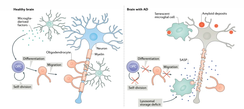
Senescence Induced Inflammation (SASP) May Promote Cell and Telomere Damage, Leading to Allodynia.

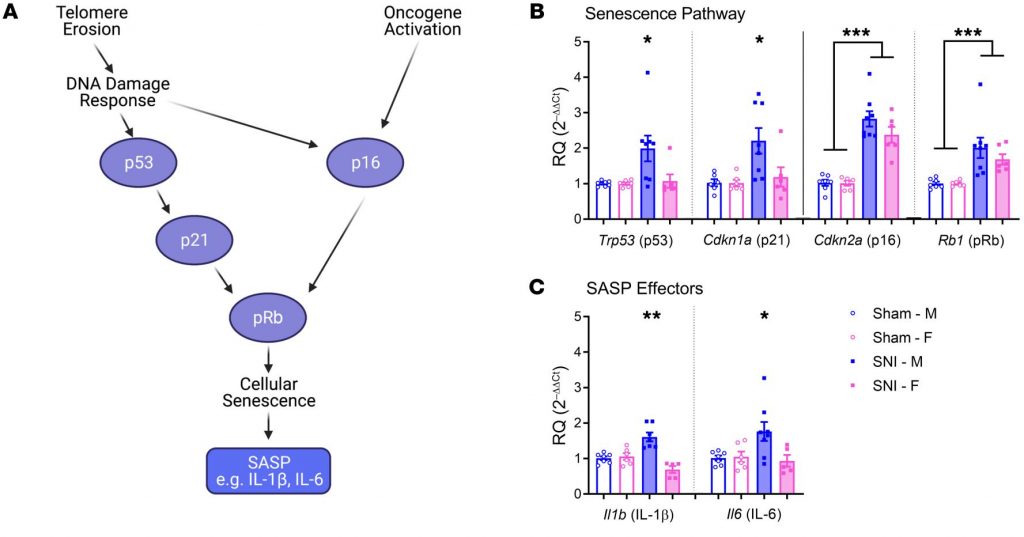
“Expression of senescence pathway and SASP effector genes in the spinal cords of mice of both sexes 6 months after sham or SNI surgery. “

(SASP is inflammation secreted by senescent cells.)
“Peripheral nerve injury produces cellular senescence in the spinal cord of mice at time points long after injury. Reduced TL can result in a persistent DNA damage response leading to cellular senescence — a state of cell cycle arrest/withdrawal, deregulated cellular metabolism, and macromolecular damage — and senescent cells in turn release a diverse set of cytokines, growth factors, proteases, and extracellular matrix components, together known as the senescence-associated secretory phenotype (SASP) or senescent-messaging secretome. Many of these SASP-related compounds are proinflammatory, and well known to produce or facilitate pain, especially when released in the spinal cord. We gave new cohorts of young male and female mice SNI or sham surgeries and harvested lumbar spinal cord tissues from these animals 12–14 months later, or 2 months later.” (1)

How can Thymosin Beta4 (TB500) improve recovery, inflammation, neuropathies, fibrosis, telomerase and senescent cell removal?

Thymosin B4 Reduces Inflammation by Upregulating MicroRNA-146a and Promotes Myelin

“Tissue inflammation results from neurological injury, and regulation of the inflammatory response is vital for neurological recovery. The innate immune response system, which includes the Toll-like receptor (TLR) proinflammatory signaling pathway, regulates tissue injury… TB4-mediated oligodendrogenesis results from [up-regulating] miR-146a [causing the] suppression [of] the TLR proinflammatory pathway and modulation of the p38 MAPK pathway.” (8)“By targeting IRAK1 and TRAF6, miR-146 inhibits NF-κB activation. We therefore hypothesized that TB4 regulates the TLR proinflammatory signaling pathway by specifically regulating miR-146a to promote differentiation of OPCs [oligodendrocyte progenitor cells] to mature myelin basic protein (MBP)-expressing OLs [oligodendrocytes]… transfection with anti-miR-146a inhibitor nucleotides significantly inhibited the expression of MBP and phosphorylation of p38 MAPK.” (8)

The Role of Senescent Cells in Alzheimer’s Disease

The Role of Senescent cells in Alzheimer’s Disease

The aging process is strongly associated with developing diseases, such as cardiovascular conditions, hypertension, cancer, diabetes mellitus, osteoporosis, and neurodegenerative diseases, among others. [10,12] Alzheimer’s disease (AD) is no exception since aging is a risk factor for late-onset AD (more than 95% of AD cases). [14,15] Another factor highly associated with aging is the increased cellular senescence population of different cell types as we approach older ages (see Fig. 3).[14] Several studies suggest cellular senescence is critical in aging and connected conditions like AD.[2,5,10] Recent investigations have pointed out that senescent cells promote the pathogenesis of AD.[3-6] But what are senescent cells? Senescent cells’ particular feature is stopping the proliferation by entering a cell cycle arrest. [12-15] these senescent cells are also known to develop apoptosis resistance and secrete proinflammatory molecules.[11] Senescent cells not only remain even though they are “damaged” but also liberate various chemicals that can initiate inflammation.[3,7] Cellular senescence emerges when a cell receives considerable stress, driving it to “reprogram” its fate to an unlimited cell cycle arrest.[7,9] DNA damage, oncogene triggering, mitochondrial dysfunction, and the accumulation of proteins like the tau and the amyloid beta (Aβ) are well known to initiate senescence in different types of cells (see Fig. 1).[2,12,14]

Figure 1. The comparison between a healthy brain and an AD brain with senescent cells.[14]

Cellular Senescence: What is it?

What is Cellular Senescence? 

Cellular senescence is a state in which cells permanently stop dividing while remaining metabolically active, typically triggered by DNA damage or other cellular stressors. First described by Leonard Hayflick in studies of human fetal fibroblasts, senescence distinguishes non-transformed cells from malignant cells, which can replicate indefinitely. Unlike quiescent cells, which can reenter the cell cycle, or terminally differentiated cells, senescent cells are permanently arrested but exhibit unique features, including chromatin reorganization, altered gene expression, and the senescence-associated secretory phenotype (SASP), a pro-inflammatory profile.The role of senescence is context-dependent, with both protective and harmful effects. It is thought to have evolved as a mechanism to prevent the malignant transformation of damaged cells. However, the accumulation of senescent cells over time contributes to age-related diseases, including cancer, tissue degeneration, and chronic inflammation. Importantly, senescence is not synonymous with aging; while aging reflects a progressive functional decline, senescence occurs throughout life and plays essential roles in embryogenesis, tissue repair, and wound healing. Despite its involvement in aging and pathology, senescence also remains a vital part of normal biological processes.

Part 2: “Cellular Senescence: What Can Be Done About It?”

Brief overview/Summary

Cellular senescence is a natural process with both protective and harmful effects. Throughout life, senescence helps prevent tumor formation and mitigate tissue damage. However, as individuals age, senescent cells accumulate in tissues, potentially contributing to various age-related diseases. Recent research has uncovered the molecular mechanisms that support the survival of senescent cells and regulate their immune clearance. These findings provide a foundation for developing new therapeutic approaches to target senescent cells while highlighting the importance of understanding the limitations, efficacy, safety, and potential risks of current strategies for senescent cell elimination. This article explores existing methods for targeting senescent cells and the challenges in advancing these strategies into safe and effective therapies. Successfully addressing these challenges could revolutionize treatments for age-related diseases and transform the way we approach health management during aging.

Peptides Targeting Mitochondrial Function and Aging.

What are mitochondria?

Mitochondria are membrane-bound cell organelles (mitochondrion, singular) that generate most of the chemical energy needed to power the cell’s biochemical reactions. Chemical energy produced by the mitochondria is stored in a small molecule called adenosine triphosphate (ATP).Mitochondria are small, often between 0.75 and 3 micrometers and are not visible under the microscope unless they are stained.
Unlike other organelles (miniature organs within the cell), they have two membranes, an outer one and an inner one. Each membrane has different functions. Mitochondria are split into different compartments or regions, each of which carries out distinct roles.Different cell types have different numbers of mitochondria. For instance, mature red blood cells have none at all, whereas liver cells can have more than 2,000. Cells with a high demand for energy tend to have greater numbers of mitochondria. Around 40 percent of the cytoplasm in heart muscle cells is taken up by mitochondria.Although mitochondria are often drawn as oval-shaped organelles, they are constantly dividing (fission) and bonding together (fusion). So, in reality, these organelles are linked together in ever-changing networks. Also, in sperm cells, the mitochondria are spiraled in the midpiece and provide energy for tail motion.

What happens to our mitochondria as we age? How do they become dysfunctional?

Age-related changes in mitochondria are associated with decline in mitochondrial function. With advanced age, mitochondrial DNA volume, integrity and functionality decrease due to accumulation of mutations and oxidative damage induced by reactive oxygen species (ROS).

In aged subjects, mitochondria are characterized by impaired function such as lowered oxidative capacity, reduced oxidative phosphorylation, decreased ATP production, significant increase in ROS generation, and diminished antioxidant defense. Mitochondrial biogenesis declines with age due to alterations in mitochondrial dynamics and inhibition of mitophagy, an autophagy process that removes dysfunctional mitochondria.

Age-dependent abnormalities in mitochondrial quality control further weaken and impair mitochondrial function. In aged tissues, enhanced mitochondria-mediated apoptosis contributes to an increase in the percentage of apoptotic cells. However, implementation of peptides and strategies such as caloric restriction and regular physical training may delay mitochondrial aging and attenuate the age-related phenotype in humans.


What is NAD+? How Does it Work?

NAD+ is the second most abundant cofactor in the human body. Anti-aging therapies are becoming more mainstream as aging is now more often being viewed as a disease. Now that this transition is happening, the ability for NAD+ to activate PARPS, Sirtuins, and help with immune dysregulation has been thoroughly investigated and NAD+ and its precursors have been highly popularized. The clinical importance of maintaining cellular NAD+ levels was established early in the last century with the finding that pellagra, a disease characterized by diarrhea, dermatitis, dementia and death, could be cured with foods containing the NAD+ precursor niacin.

Additionally, cellular concentrations of NAD+ have been shown to decrease under conditions of increased oxidative damage such as occur during aging Altered levels of NAD+ have been found to accompany several disorders associated with increased oxidative/free radical damage including diabetes, heart disease, age-related vascular dysfunction, ischemic brain injury, misfolded neuronal proteins, and Alzheimer’s dementia. Interventions targeted at restoring NAD+ has been shown in animal models to support healthy aging and improve metabolic function, and dementia.

A need for NAD+ in muscle development, homeostasis, and aging

In a review study, researchers discuss the recent data that document conserved roles for NAD+ in skeletal muscle development, regeneration, aging, and disease as well as interventions targeting skeletal muscle and affecting NAD+ that suggest promising therapeutic benefits. The researchers also highlight gaps in our knowledge and propose avenues of future investigation to better understand why and how NAD+ regulates skeletal muscle biology.

Peptides Targeting Mitochondrial Function and Aging.

What are mitochondria?

Mitochondria are membrane-bound cell organelles (mitochondrion, singular) that generate most of the chemical energy needed to power the cell’s biochemical reactions. Chemical energy produced by the mitochondria is stored in a small molecule called adenosine triphosphate (ATP).Mitochondria are small, often between 0.75 and 3 micrometers and are not visible under the microscope unless they are stained.
Unlike other organelles (miniature organs within the cell), they have two membranes, an outer one and an inner one. Each membrane has different functions. Mitochondria are split into different compartments or regions, each of which carries out distinct roles.Different cell types have different numbers of mitochondria. For instance, mature red blood cells have none at all, whereas liver cells can have more than 2,000. Cells with a high demand for energy tend to have greater numbers of mitochondria. Around 40 percent of the cytoplasm in heart muscle cells is taken up by mitochondria.Although mitochondria are often drawn as oval-shaped organelles, they are constantly dividing (fission) and bonding together (fusion). So, in reality, these organelles are linked together in ever-changing networks. Also, in sperm cells, the mitochondria are spiraled in the midpiece and provide energy for tail motion.


What happens to our mitochondria as we age? How do they become dysfunctional?

Age-related changes in mitochondria are associated with decline in mitochondrial function. With advanced age, mitochondrial DNA volume, integrity and functionality decrease due to accumulation of mutations and oxidative damage induced by reactive oxygen species (ROS).

In aged subjects, mitochondria are characterized by impaired function such as lowered oxidative capacity, reduced oxidative phosphorylation, decreased ATP production, significant increase in ROS generation, and diminished antioxidant defense. Mitochondrial biogenesis declines with age due to alterations in mitochondrial dynamics and inhibition of mitophagy, an autophagy process that removes dysfunctional mitochondria.

Age-dependent abnormalities in mitochondrial quality control further weaken and impair mitochondrial function. In aged tissues, enhanced mitochondria-mediated apoptosis contributes to an increase in the percentage of apoptotic cells. However, implementation of peptides and strategies such as caloric restriction and regular physical training may delay mitochondrial aging and attenuate the age-related phenotype in humans.

Anti-Inflammatory and Anti-Fibrotic Effects of Thymosin Beta4 Fragment Ac-SDKP.

Ac-SDKP is a Peptide Fragment of Thymosin Beta 4 (TB-500)

“Tβ4 is a naturally occurring peptide consisting of 43 base pairs of amino acids and generates the N-terminal tetrapeptide AcSDKP.” (14)

Peptide Ac-SDKP was isolated from the whole Thymosin Beta 4 Peptide Sequence.

Ac-SDKP Reduced Kidney Fibrosis.

“Treatment of cultured cells with ACEi alone or in combination with AcSDKP prevented the downregulated expression of miR-29s and miR-let-7s induced by TGFβ stimulation. Interestingly, ACEi also restored miR-29 and miR-let-7 family cross-talk in endothelial cells, an effect that is shared by AcSDKP suggesting that AcSDKP may be partially involved in the anti-mesenchymal action of ACEi. The results of the present study promise to advance our understanding of how ACEi regulates antifibrotic microRNAs crosstalk and DPP-4 associated-fibrogenic processes which is a critical event in the development of diabetic kidney disease.” (14)

What is Thymosin Beta 4, How Does it Work?

What is Thymosin Beta-4?

The beta-thymosins (b-thymosins) comprise a family of structurally related, highly conserved amino acid sequences in species ranging from mammals to echinoderms. Of the 16 known family members, thymosin β4 (Tb4), thymosin β10 (Tb10), and thymosin β15 (Tb15) are found in man.Thymosin beta-4 (TB4) is a 43 amino acid, 5kDa polypeptide that is an important mediator of cell proliferation, migration, and differentiation. TB4 is the most abundant member of the β- thymosin family in mammalian tissue and is regarded as the main G-actin sequestering peptide. It is found in all tissues and cell types except red blood cells. Thymosin beta-4 is angiogenic and can promote endothelial cell migration and adhesion, and angiogenesis. TB4 also accelerates wound healing and reduces inflammation and scarring when applied in dermal wound-healing assays.

Beta thymosins bind and sequester monomeric actin, thus preventing actin polymerization and formation of filamentous actin. Actin is a vital component of cell structure and movement. Actin is involved in many important non-muscle cellular processes, including cell locomotion, chemotaxis, phagocytosis, and cytokinesis. Of the thousands of proteins present in cells, actin makes up to 10% of the total proteins in a cell, representing a major role in the genetic makeup of the cell.

Animal studies of disease and repair when using thymosin beta-4, the major actin-sequestering molecule in mammalian cells, have provided a base for the ongoing multicenter clinical trials for wound healing, including dermal, corneal, and cardiac. TB4 has multiple biological activities, which include down-regulation of inflammatory chemokines and cytokines, and promotion of cell migration, blood vessel formation, cell survival, and stem cell maturation.

Thymosin beta-4 also inhibits inflammation, microbial growth, scar formation (by reducing the level of myofibroblasts), and apoptosis, and protects cells from cytotoxic damage, including glutamate neuronal toxicity. In addition, it binds to G-actin, blocks actin polymerization, and is released with factor X by platelets. These activities contribute to the multiple wound healing properties that have been reported in animal and human studies.

Welcome back!

or
Menu
Explore

Search Peptides

Welcome back!

or
0