Research - Page 14 of 16 - Ai-Peptides

FREE SHIPPING ON ORDERS OVER $100

Research Categories
[list_research_categories]
Research Categories

Research

ALL ARTICLES AND PRODUCT INFORMATION PROVIDED ON THIS WEBSITE ARE FOR INFORMATIONAL AND EDUCATIONAL PURPOSES ONLY. The products offered on this website are furnished for in-vitro studies only. In-vitro studies (Latin: in glass) are performed outside of the body. These products are not medicines or drugs and have not been approved by the FDA to prevent, treat or cure any medical condition, ailment or disease. Bodily introduction of any kind into humans or animals is strictly forbidden by law.

How Do Telomeres Impact Aging and Autophagy?

Loss of Telomere Leads to p53 and Autophagy Induced Cell Death.

“autophagy-deficient cells … continued to proliferate and bypassed crisis” (2)
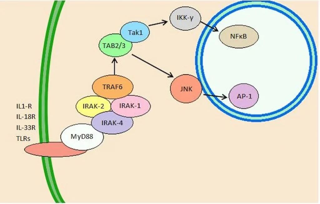
“In summary, cells in telomere crisis undergo cell death through autophagy, which is triggered by chromosome breakage and transduced by the cGAS–STING pathway. As cell death in crisis represents the final barrier against neoplastic transformation, a cancer therapy that involves inhibition of autophagy could be counterproductive… Moreover, cells lacking either cGAS or STING proliferated beyond crisis. “”Autophagy mediates the turnover of cytoplasmic macromolecules to support cellular homeostasis. Autophagy generally blocks apoptosis, but in specific circumstances it can lead to cell death through excessive degradation of cell constituents. The authors studied telomere crisis using human fibroblasts and epithelial cells, in which the RB and/or p53 pathways were suppressed; these cells bypassed senescence and entered replicative stress, exhibiting telomere attrition, chromosome fusions and cell death.””Telomere deprotection through TRF2 depletion was sufficient to activate autophagy independently of replicative crisis, and genetic suppression of telomere fusions in TRF2-depleted cells reduced the accumulation of cytosolic DNA and attenuated autophagy, suggesting that fusion-dependent cytosolic DNA is required for the telomeric autophagy response. ” (2)

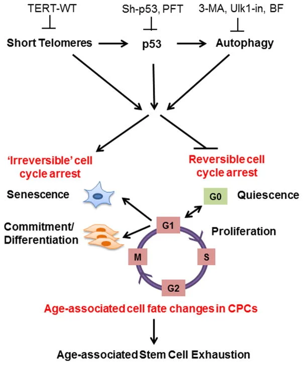
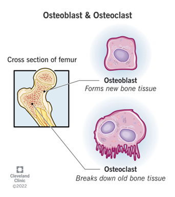
“The cell fate of CPCs changes with age and is characterized by a switch away from proliferation and quiescence (reversible form of cell cycle arrest) towards senescence and increased basal commitment (‘irreversible’ forms of cell cycle arrest) accounting for age-associated stem cell exhaustion. Mechanistically, short telomeres activate p53 that induces autophagy and at least partially contributes to the age-associated change in cell fate. Blunting telomere shortening via overexpression of TERT-WT, silencing p53 , or treating with pharmacological inhibitors of p53 (PFT) and autophagy (3-MA, Ulk1-In, BF) selectively attenuate senescence and basal commitment and reverse cell fate of aged CPCs.” (3)

Circadian Rhythm Controls Telomeres and Telomerase Activity.

“Circadian clocks are fundamental machinery in organisms ranging from archaea to humans. Disruption of the circadian system is associated with premature aging in mice, but the molecular basis underlying this phenomenon is still unclear. In this study, we found that telomerase activity exhibits endogenous circadian rhythmicity in humans and mice. Human and mouse TERT mRNA expression oscillates with circadian rhythms and are under the control of CLOCK–BMAL1 heterodimers.

CLOCK deficiency in mice causes loss of rhythmic telomerase activities, TERT mRNA oscillation, and shortened telomere length. Physicians with regular work schedules have circadian oscillation of telomerase activity while emergency physicians working in shifts lose the circadian rhythms of telomerase activity. These findings identify the circadian rhythm as a mechanism underlying telomere and telomerase activity control that serve as interconnections between circadian systems and aging.” (4)

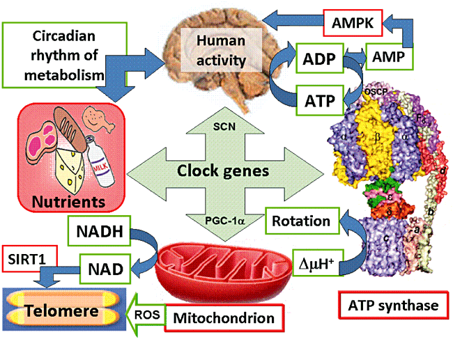
“Human activity is driven by NADH and ATP produced from nutrients, and the resulting NAD and AMP play a predominant role in energy regulation. Caloric restriction increases both AMP and NAD and is known to extend the healthspan (healthy lifespan) of animals. Silent information regulator T1 (SIRT1), the NAD-dependent deacetylase, attenuates telomere shortening, while peroxisome proliferator-activated receptor γ coactivator 1α (PGC-1α), a master modulator of gene expression, is phosphorylated by AMP kinase and deacetylated by SIRT1. Thus, PGC-1α is a key component of the circadian oscillator that integrates the mammalian clock and energy metabolism.

Reactive oxygen species produced in clock mutants result in telomere shortening. The circadian rhythms produced by clock genes and lifestyle factors are ultimately controlled by the human brain and drive homeostatic and hedonic feeding and daily activity. ” (9).

  • Telomerase and TERT mRNA expressions exhibit endogenous circadian rhythm.
  • Human and mouse TERT mRNA expression are under the control of CLOCK–BMAL1 heterodimers.
  • CLOCK deficient mice have shortened telomere length and abnormal oscillations of telomerase activity and TERT mRNA.
  • Emergency physicians working in shifts lose the circadian rhythms of telomerase activity.

TB-500 (Thymosin Beta-4) Peptide Research

TB-500 also known as Thymosin Beta 4 is a naturally occurring peptide. It is found in high concentrations in blood platelets, wound fluid and other tissues in the body. TB-500 is not a growth factor; rather, it is a major actin regulating peptide. TB-500 (Thymosin Beta 4) has been found to play an important role in protection, regeneration and remodeling of injured or damaged tissues. The gene for TB-500 (Thymosin Beta 4) has also been found to be one of the first to be upregulated after a wound occurs.

Thymosin Beta 4 and Cardiac Regeneration

Thymosin β4 and Prothymosin α Promote Cardiac Regeneration Post-Ischaemic Injury in Mice

“The adult mammalian heart is a post-mitotic organ. Even in response to necrotic injuries, where regeneration would be essential to reinstate cardiac structure and function, only a minor percentage of cardiomyocytes undergo cytokinesis… In this study, we aimed to determine the gene expression profile of proliferating adult cardiomyocytes in the mammalian heart after myocardial ischaemia, to identify factors to can promote cardiac regeneration… Here, we demonstrate increased 5-ethynyl-2’deoxyuridine incorporation in cardiomyocytes 3 days post-myocardial infarction in mice… Combinatorial overexpression of the enriched genes within this population in neonatal rat cardiomyocytes and mice at postnatal day 12 (P12) unveiled key genes that promoted increased cardiomyocyte proliferation. Therapeutic delivery of these gene cocktails into the myocardial wall after ischaemic injury demonstrated that a combination of thymosin beta 4 (TMSB4) and prothymosin alpha (PTMA) provide a permissive environment for cardiomyocyte proliferation and thereby attenuated cardiac dysfunction… This study reveals the transcriptional profile of proliferating cardiomyocytes in the ischaemic heart and shows that overexpression of the two identified factors, TMSB4 and PTMA, can promote cardiac regeneration.” (2)

The Role of Thymosin β4 in Cardiomyocyte Proliferation and Cardiac Regeneration

“Thymosin β4 (Tβ4) is a 43-amino acid protein that belongs to the β-thymosin family, which is highly conserved… Tβ4 has been associated with wound healing, inflammation, fibrosis, and tissue regeneration, with recent studies suggesting that Tβ4 can help prevent inflammation and fibrosis in the eye, skin, lung, and liver… Tβ4 is a potent protective factor that can protect against myocyte damage, promote myocyte regeneration, and inhibit heart inflammation… Therefore, we propose that Tβ4 might have an antifibrotic function in the heart.” (3)

How can Thymosin Beta4 (TB500) improve recovery, inflammation, neuropathies, fibrosis, telomerase and senescent cell removal?

Thymosin B4 Reduces Inflammation by Upregulating MicroRNA-146a and Promotes Myelin

“Tissue inflammation results from neurological injury, and regulation of the inflammatory response is vital for neurological recovery. The innate immune response system, which includes the Toll-like receptor (TLR) proinflammatory signaling pathway, regulates tissue injury… TB4-mediated oligodendrogenesis results from [up-regulating] miR-146a [causing the] suppression [of] the TLR proinflammatory pathway and modulation of the p38 MAPK pathway.” (8)“By targeting IRAK1 and TRAF6, miR-146 inhibits NF-κB activation. We therefore hypothesized that TB4 regulates the TLR proinflammatory signaling pathway by specifically regulating miR-146a to promote differentiation of OPCs [oligodendrocyte progenitor cells] to mature myelin basic protein (MBP)-expressing OLs [oligodendrocytes]… transfection with anti-miR-146a inhibitor nucleotides significantly inhibited the expression of MBP and phosphorylation of p38 MAPK.” (8)

Cagrilintide Research: What is Cagrilintide? How does it work?

The Obesity Epidemic

Obesity has become one of the most pressing public health challenges in the United States and around the world. In the U.S., about 42% of adults are classified as obese, a figure that has nearly tripled over the past 50 years. Globally, over 650 million people are classified as obese, and this number continues to rise rapidly. Obesity increases the risk for many serious health conditions, including heart disease, type 2 diabetes, stroke, and certain cancers, making it a major contributor to premature death and disability worldwide.

Obesity is difficult to control due to several complex factors. It’s not simply a matter of overeating or lack of exercise; rather, it stems from a mix of genetics, hormonal imbalances, metabolism, and environmental factors, such as the easy availability of high-calorie foods and increasingly sedentary lifestyles. This complexity makes it challenging to address basic “eat less, move more” strategies. Additionally, many individuals struggle with yo-yo dieting, where initial weight loss is followed by weight regain once normal eating patterns resume. This cycle makes maintaining weight loss over the long term even more difficult. Furthermore, the medications currently available for weight loss often have limited effectiveness and can come with side effects, making them less appealing for long-term use. Most of these drugs only target specific aspects of obesity, like appetite suppression or metabolism, which doesn’t fully address the root causes of the issue.

Peptides BPC157, AOD9604, MOTS-c improve Bone Mineral Density for Osteoporosis.

Osteoporosis is the most prevalent systemic skeletal system disease, leading to increased bone fragility and vulnerability to fractures. Due to the microarchitectural destruction in bone tissue, fracture healing in osteoporoti patients is often delayed and compromised compared with non‑osteoporotic individuals. Osteoporosis usually results from meno‑ pause, aging, metabolic diseases and drug therapies with the precise cellular and molecular mechanism remaining to be elucidated.

Recent studies have shown that four peptides (BPC-157AOD 9604MOTS-c, Peptide 11R‐VIVIT) have been proven to have healing effects for such disease in several types of model… High concentration and long-term stimulation of TGF-β1 induced osteogenic differentiation of bone marrow mesenchymal stem cells (MSCs) in vitr2. TGF-β pathway-related genes exert anti-osteoporosis effects by regulating the function of bone deposits and osteoclasts. TGF-β also affects the bone formation by promoting the proliferation and differentiation of osteoblasts, as well as the synthesis of extracellular matrix.

Peptides for Bone Healing Research

Our bones are the foundation of our bodies, protecting vital organs like the brain and heart while providing a solid structure against which muscles can work. They are also responsible for producing most of our red blood cells and play a critical role in the immune system. Bone problems are becoming more prevalent, however, and this has led researchers to investigate potential means of mitigating these problems. Peptides have emerged as one of the forerunners in the fight to preserve bone health with peptides like ipamorelin, transforming-growth factor-beta, bone morphogenic proteins, and others showing a great deal of promise.

Increasing Prevalence of Bone Problems

Despite the common perception of bones as static, and unchanging structures, research has shown them to be exceptionally dynamic. Bone isn’t simply deposited as we grow and then stops, but rather is in a constant state of flux. The balance between bone growth and bone breakdown is necessary to maintain blood calcium levels, allow bones to respond to changes in stressors, promote bone healing following injury, and much more. As it turns out, diseases that affect bone are actually diseases that alter this careful balance and thus treating them is more complex than simply encouraging bones to grow.

Bone deterioration and disease are becoming more and more commonplace for several reasons. First, as the population ages the incidence of bone-related diseases like osteoporosis is increasing as well[1]. Aging is the single greatest risk factor for bone loss, but it isn’t the only contributor to the problem. Other reasons for increased bone disease include vitamin D deficiency, increased prevalence of diseases that impact bone health, increasing use of medications that degrade bone health, nutritional issues, and increasingly sedentary lifestyles.

Of course, bone loss and degradation are not the only problems to confront our skeletal system. Fractures that result from trauma are also a major cause of lost time at work as well as lost quality time. Many fractures take eight to twelve weeks to heal, so anything that can be done to speed up this recovery process would help to drive down both societal and medical costs.

PEPITEM Research: Osteoporosis and Bone Strength

PEPITEM (Peptide Inhibitor of Trans-Endothelial Migration)

PEPITEM (Peptide Inhibitor of Trans-Endothelial Migration) was first identified in 2015 by researchers at the University of Birmingham. New research published in Cell Reports Medicine demonstrates for the first time that PEPITEM could be utilized as a novel early clinical intervention to mitigate the effects of age-related musculoskeletal diseases. The data indicates that PEPITEM enhances bone mineralization, formation, and strength, effectively reversing bone loss in animal models.

This research was supported by significant grants from the Medical Research Council and the Lorna and Yuti Chernajovsky Biomedical Research Foundation, which invests in research for developing new targeted medicines to improve health outcomes. Additional funding was provided by the British Society for Research on Ageing and Versus Arthritis.

Bone undergoes constant formation, reformation, and remodeling throughout life, with up to 10% of human bone being replaced annually. This process involves a complex interplay between osteoblasts, which form bone, and osteoclasts, which break down bone. Disruptions to this finely tuned process are responsible for diseases such as osteoporosis and rheumatoid arthritis, characterized by excessive bone breakdown, or ankylosing spondylitis, marked by abnormal bone growth.

PEPITEM’s ability to enhance bone formation without adversely affecting osteoclast activity presents a promising therapeutic strategy. By promoting osteoblast activity and reducing osteoclast numbers via specific signaling pathways, PEPITEM has shown potential in increasing bone density and strength, suggesting its effectiveness as an early intervention for age-related bone diseases. This advancement could offer a new approach to maintaining bone health and preventing fractures, addressing a significant need in the treatment of osteoporosis and other musculoskeletal disorders.

Current osteoporosis therapies, such as bisphosphonates, primarily target osteoclasts to prevent further bone loss. While there are new anabolic agents available that promote new bone formation, their clinical use is limited. For example, teriparatide (parathyroid hormone, or PTH) is effective for only 24 months, and romosozumab (an anti-sclerostin antibody) has been linked to cardiovascular events. These limitations show the urgent need for developing new therapies to stimulate bone repair, especially in age-related musculoskeletal diseases like osteoporosis.

To address this need, a team of researchers led by Dr. Helen McGettrick and Dr. Amy Naylor, along with Dr. Jonathan Lewis and Ms. Kathryn Frost from the Institute of Inflammation and Ageing at the University of Birmingham, and Dr. James Edwards from the Nuffield Department of Orthopedics, Rheumatology and Musculoskeletal Sciences at the University of Oxford, have been investigating the therapeutic potential of PEPITEM (Peptide Inhibitor of Trans-Endothelial Migration) in these conditions. Their research aims to explore whether PEPITEM can effectively stimulate bone repair and provide a new therapeutic avenue for treating osteoporosis and other age-related musculoskeletal disorders. By enhancing bone mineralization, formation, and strength, PEPITEM offers an exciting alternative that could overcome the limitations of existing therapies and significantly improve patient outcomes.

The Role of Cell-Permeable Peptides and The JNK Family in Preventing Neuronal Degeneration

Figure 1:     Neuroprotective features of CPPs in combination with other molecules or peptides for treating PD and AD.

JNK family participates in the apoptosis pathway, also known as programmed cell death.[4] The apoptosis mechanism is used to eliminate cells that are damaged in anyway. This mechanism is suitable for healthy people because it helps avoid developing cancer and other disorders.[1] On the other hand, apoptosis is detrimental in neurodegenerative diseases that share the abnormal accumulation of misfolded proteins, contributing to dementia, cognitive loss, memory loss, behavioral problems, and sleep problems, among others, through neuronal cell loss.[3,5,7] In addition, the JNK family also participates in pathways related to regulation, plasticity, development of the Central Nervous System (CNS), inflammation, and autophagy.[9] The well-functioning of all these processes is vital. Recent studies suggested that an imbalance of the JNK family members can accelerate the progression of both AD and PD pathologies. One of the hallmarks of AD is the aggregation of amyloid beta (Aβ) in the extracellular area.[7] The overexpression of Aβ triggers the activation of JNK-3, which also participates in the formation of Aβ42, a toxic species that affects the neuron’s cell function when it accumulates.[7] It is unclear whether the neurodegenerative disorder or the JNK imbalance comes first. However, they both have a direct relationship where PD and AD patients show high levels of JNK in the brain (postmortem), and irregular levels of the JNK family accelerate the progression of both disorders (see Fig. 2). [5,9] JNK imbalance could also lead to increased inflammation responses, low plasticity, and a flaw in autophagy performance, among other related adverse effects. An increase in apoptosis in PD and AD patients causes a decrease in neurons in the brain, affecting cognitive, behavioral, and memory performance.[8] JNK also decreases the α-syn accumulation in PD models, contributing to neuroprotection.[10] JNK pathway influence positively many vital processes of the body unless an imbalance occurs. [2,8] JNK has been the target for treating several diseases like cancer, strokes, PD, AD, Huntington’s disease, and other neurodegenerative disorders, showing promising results. More investigations need to be done to comprehend better how the correct levels of JNK can help combat important hallmarks of degenerative illnesses to eventually prevent or revert the progression of related diseases like PD and AD. In general, the benefits of inhibiting JNKs for treating neurodegenerative disorders are:

­Improving autophagy for the elimination of misfolded proteins (Aβ (AD), tau protein (AD), and α-syn (PD)

  • Increasing cell proliferation.
  • ­Decreasing programmed cell death.­
  • Reducing brain damage.
  • Decreasing cognitive decline.
  • Increasing gene expression.
  • Decreasing neuroinflammation responses
  • Improving memory
  • Providing neuroprotection
  • Improving synaptic connections

Amylin Protein Targets Alzheimer’s disease

The remarkable benefits of amylin protein as a treatment for Alzheimer’s disease

Alzheimer’s disease (AD) is known to be a multifactorial disease. [2] That is, several factors contribute to the pathogenic development of this disorder. However, amyloid beta () accumulation in the brain is highlighted as critical in developing the disease. [2,3] Recent studies have shown that long before the onset of symptoms of the disease, the accumulation of the Aβ protein forms plaques between neurons which turn out to be very toxic to neurons. [2,3] In addition, it has been exposed that these plaques are responsible for progressive cognitive impairment. [1,2,3] The accumulation of Aβ induces neuronal cells to die. Aβ plaques constantly induce neuroinflammation, so it, in turn, causes injury to nerve cells affecting memory and other cognitive factors. [2,3]

Representation of the hallmarks of AD in the brain.

Countless studies have found a protein with Aβ like characteristics called amylin[1,2,3,5] These two proteins generate soluble forms of oligomeric form intermediates, which have been found to have potent cytotoxicity. [2,6] This cytotoxicity affects cell membranes and organelles, inducing inflammatory responses, causing reactive oxygen species, and overloading the protein-splitting. [1,2] These two proteins are so similar that their oligomers can even interact with each other accumulating in the brain in the same way as Aβ alone. [1,2] Amylin accumulation in the brain of AD rat models has been found to contribute significantly to brain damage caused by the illness. [2,6]In addition to AD, amylin has been found in high concentrations in the brain of patients with dementia and type 2 diabetes(T2DM). [2]

Welcome back!

or
Menu
Explore

Search Peptides

Welcome back!

or
0