FREE SHIPPING ON ORDERS OVER $100

Research Categories
[list_research_categories]
Research Categories

Research

ALL ARTICLES AND PRODUCT INFORMATION PROVIDED ON THIS WEBSITE ARE FOR INFORMATIONAL AND EDUCATIONAL PURPOSES ONLY. The products offered on this website are furnished for in-vitro studies only. In-vitro studies (Latin: in glass) are performed outside of the body. These products are not medicines or drugs and have not been approved by the FDA to prevent, treat or cure any medical condition, ailment or disease. Bodily introduction of any kind into humans or animals is strictly forbidden by law.

Telomeres and Epithalon

Telomeres & Epithalon

Introduction to Telomeres

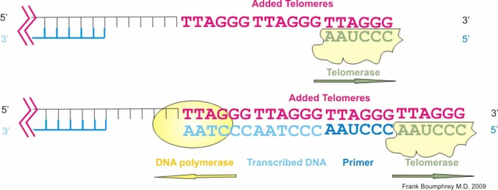
Telomeres are repeating sequences of nucleotide sequences (TTAGGG) that tag the ends of all chromosomes. They are designed to prevent unpredictable changes in the DNA strand, keeping the genome stable.

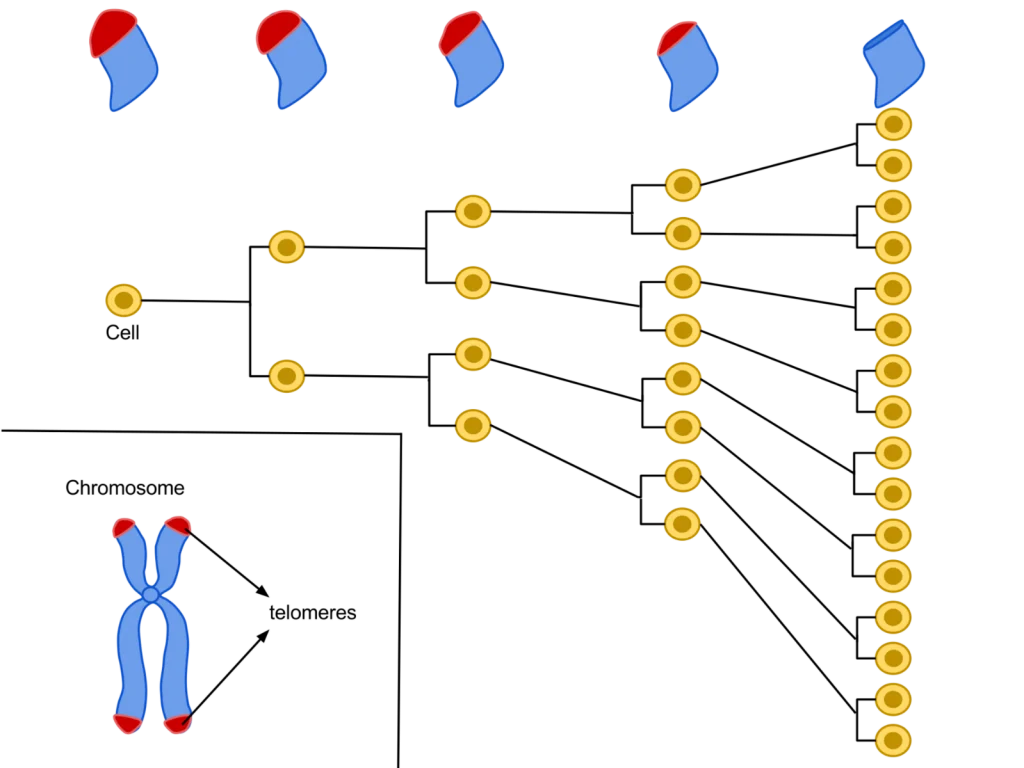
Their primary function is to prevent chromosomal “fraying” when a cell replicates, much like the plastic tips on the end of shoelaces. As a cell ages, its telomeres become shorter.

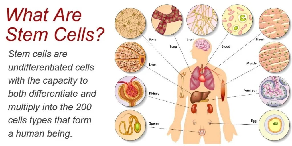
This shortening is thought to be one of several factors that causes cells to age. In actively dividing cells, such as those in the bone marrow, the stem cells of the embryo, and germ cells in the adult, telomere length (TL) is kept constant by the enzyme telomerase.

As the organism grows, this enzyme becomes less active over time. This leads to a slow decrease in telomere length, until a point is reached at which the cell is no longer capable of replication (‘replicative senescence’). A cell can no longer divide when telomeres are too short—once they reach a critical point, the cell becomes inactive (or ‘senescent’), slowly accumulating damage that it can’t repair, or it dies.

What is Epithalon?

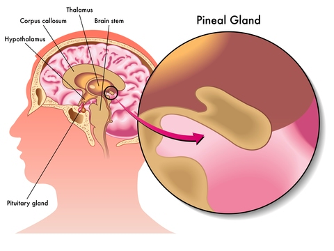
Epithalon (aka Epitalon) peptide (Ala- Glu-Asp-Gly) was constructed and synthesized based on amino acid composition of Epithalamine, a complex peptide preparation isolated from animal brain pineal. It was first discovered in the late 1980’s by Prof. Vladimir Khavinson from The Sankt Petersburg University, Russia.

As the most prominent tasks of the pineal gland are to maintain different kind of processes in our body, such as to normalize the activity of anterior pituitary and to maintain the levels of calcium, gonadotropins, and melatonin, its activity is highly regulated by a series of feedback mechanisms. Epithalamin acts as an antioxidant and increases the resistance to stress and lowers the levels of corticosteroids. The life extension and anti-aging properties, amongst a variety of different clinical indications, of epithalon are incredible antioxidant and increases the resistance to stress and lowers the levels of corticosteroids. The life extension and anti-aging properties, amongst a variety of different clinical indications, of epithalon are incredible.

What is Epithalon and How Does it Protect Telomeres?

What is Epithalon?

Epithalon is a short, 4 amino acid chain peptide used to regulate the cell cycle through the upregulation of telomerase activity. It has been shown to have distinctive anti-aging and anti-tumor activity across many animal and human studies. Known as the synthetic version of the tetrapeptide epithalamin, which naturally occurs in the pineal gland in our body, Epithalon (also known as Epitalon or Epithalone) was first discovered in the late 1980’s by Prof. Vladimir Khavinson from The Sankt Petersburg University, Russia.

As the most prominent tasks of the pineal gland are to maintain different kind of processes in our body, such as to normalize the activity of anterior pituitary and to maintain the levels of calcium, gonadotropins, and melatonin, its activity is highly regulated by a series of feedback mechanisms. Epithalamin acts as an antioxidant and increases the resistance to stress and lowers the levels of corticosteroids. The life extension and anti-aging properties, amongst a variety of different clinical indications, of epithalon are incredible.

Scientific research has revealed that epithalon affects the following:

  • Upregulate telomerase activity
  • Normalize antioxidant indices
  • Reduce peroxide lipid oxidation products
  • Increase activity of glutathione peroxidase
  • Improve melatonin and immunity (cellular and humoral)
  • Improve insulin sensitivity
  • Decrease LDL and VLDL
  • Improve tissue repair
  • Anti-tumor effects
  • Decrease mortality and increases life expectancy


Epithalon Fights Aging by Activating Telomerase

Epithalon, also known as Epitalon is a synthetic peptide analog of epithalamin, a protein found in the pineal gland of mammals and of interest for its anti-aging properties. Past research studies have demonstrated that epithalamin can increase maximum life span in animals, decrease levels of free radicals, and alter catalase activity to prevent tissue damage [1]. Epithalamin has been shown to decrease mortality by 52% in fruit flies, by 52% in normal rats, and by 27% in mice prone to certain types of cancer and cardiovascular disease [2].

It has been shown, through extensive animal research, to be a potent regulator of cell metabolism, including growth and cell division. In particular, epithalon is able to extend cell survival in vitro. At least part of the reason that epithalon can extend cell survival comes down to its action on telomeres.

Epithalon has similar effects to epithalamin in mice and rats. It has also shown promise as an anti-cancer agent, reducing spontaneous mammary tumors in mice prone to them and reducing incidence of intestinal tumors in rodents. How does it achieve these effects?

What is Epithalon?

Epithalon is currently being studied and researched by Scientists and Doctors specializing in the field of anti-aging care and medicine. EPITHALON (Epitalon) is one of the most important breakthroughs in the study of anti-aging.

The Epithalon (Epitalon) tetrapeptide has been discovered by researchers in Russia. This was seen to reactivate the production of cell telomerase thus slowing down the aging process and rejuvenating the entire body. The development of molecular biology required bio-chemical studies that were nothing short of profound. Scientific work by Gobind Khorana and Marshall Nirenberg for many years resulted in defining codons or nucleotides and triplets and the genetic code of each of the 20 amino acids. This resulted in a Nobel Prize award in 1968 with Robert Holley. Nucleic acid investigations and identification of DNA and RNA base sequences were also conducted by the 1980 Nobel Prize winner for Chemistry Frederick Sanger along with Walter Gilbert and Paul Berg. These studies revealed the cause of aging.

How Do Telomeres Impact Aging and Autophagy?

Loss of Telomere Leads to p53 and Autophagy Induced Cell Death.

“autophagy-deficient cells … continued to proliferate and bypassed crisis” (2)
“In summary, cells in telomere crisis undergo cell death through autophagy, which is triggered by chromosome breakage and transduced by the cGAS–STING pathway. As cell death in crisis represents the final barrier against neoplastic transformation, a cancer therapy that involves inhibition of autophagy could be counterproductive… Moreover, cells lacking either cGAS or STING proliferated beyond crisis. “”Autophagy mediates the turnover of cytoplasmic macromolecules to support cellular homeostasis. Autophagy generally blocks apoptosis, but in specific circumstances it can lead to cell death through excessive degradation of cell constituents. The authors studied telomere crisis using human fibroblasts and epithelial cells, in which the RB and/or p53 pathways were suppressed; these cells bypassed senescence and entered replicative stress, exhibiting telomere attrition, chromosome fusions and cell death.””Telomere deprotection through TRF2 depletion was sufficient to activate autophagy independently of replicative crisis, and genetic suppression of telomere fusions in TRF2-depleted cells reduced the accumulation of cytosolic DNA and attenuated autophagy, suggesting that fusion-dependent cytosolic DNA is required for the telomeric autophagy response. ” (2)

“The cell fate of CPCs changes with age and is characterized by a switch away from proliferation and quiescence (reversible form of cell cycle arrest) towards senescence and increased basal commitment (‘irreversible’ forms of cell cycle arrest) accounting for age-associated stem cell exhaustion. Mechanistically, short telomeres activate p53 that induces autophagy and at least partially contributes to the age-associated change in cell fate. Blunting telomere shortening via overexpression of TERT-WT, silencing p53 , or treating with pharmacological inhibitors of p53 (PFT) and autophagy (3-MA, Ulk1-In, BF) selectively attenuate senescence and basal commitment and reverse cell fate of aged CPCs.” (3)

Circadian Rhythm Controls Telomeres and Telomerase Activity.
“Circadian clocks are fundamental machinery in organisms ranging from archaea to humans. Disruption of the circadian system is associated with premature aging in mice, but the molecular basis underlying this phenomenon is still unclear. In this study, we found that telomerase activity exhibits endogenous circadian rhythmicity in humans and mice. Human and mouse TERT mRNA expression oscillates with circadian rhythms and are under the control of CLOCK–BMAL1 heterodimers.

CLOCK deficiency in mice causes loss of rhythmic telomerase activities, TERT mRNA oscillation, and shortened telomere length. Physicians with regular work schedules have circadian oscillation of telomerase activity while emergency physicians working in shifts lose the circadian rhythms of telomerase activity. These findings identify the circadian rhythm as a mechanism underlying telomere and telomerase activity control that serve as interconnections between circadian systems and aging.” (4)

“Human activity is driven by NADH and ATP produced from nutrients, and the resulting NAD and AMP play a predominant role in energy regulation. Caloric restriction increases both AMP and NAD and is known to extend the healthspan (healthy lifespan) of animals. Silent information regulator T1 (SIRT1), the NAD-dependent deacetylase, attenuates telomere shortening, while peroxisome proliferator-activated receptor γ coactivator 1α (PGC-1α), a master modulator of gene expression, is phosphorylated by AMP kinase and deacetylated by SIRT1. Thus, PGC-1α is a key component of the circadian oscillator that integrates the mammalian clock and energy metabolism.

Reactive oxygen species produced in clock mutants result in telomere shortening. The circadian rhythms produced by clock genes and lifestyle factors are ultimately controlled by the human brain and drive homeostatic and hedonic feeding and daily activity. ” (9)

  • Telomerase and TERT mRNA expressions exhibit endogenous circadian rhythm.
  • Human and mouse TERT mRNA expression are under the control of CLOCK–BMAL1 heterodimers.
  • CLOCK deficient mice have shortened telomere length and abnormal oscillations of telomerase activity and TERT mRNA.
  • Emergency physicians working in shifts lose the circadian rhythms of telomerase activity.

Cellular Senescence and Chronic Pain

Senescence Induced Inflammation (SASP) May Promote Cell and Telomere Damage, Leading to Allodynia.

“Expression of senescence pathway and SASP effector genes in the spinal cords of mice of both sexes 6 months after sham or SNI surgery. “

(SASP is inflammation secreted by senescent cells.)

“Peripheral nerve injury produces cellular senescence in the spinal cord of mice at time points long after injury. Reduced TL can result in a persistent DNA damage response leading to cellular senescence — a state of cell cycle arrest/withdrawal, deregulated cellular metabolism, and macromolecular damage — and senescent cells in turn release a diverse set of cytokines, growth factors, proteases, and extracellular matrix components, together known as the senescence-associated secretory phenotype (SASP) or senescent-messaging secretome. Many of these SASP-related compounds are proinflammatory, and well known to produce or facilitate pain, especially when released in the spinal cord. We gave new cohorts of young male and female mice SNI or sham surgeries and harvested lumbar spinal cord tissues from these animals 12–14 months later, or 2 months later.” (1)

Posted in:

How does Epitalon enhance sleep while protecting DNA and Telomeres?

Epithalon Peptide Induces Telomerase Activity and Telomere Elongation in Human Somatic Cells and Overcomes the Hayflick Limit

Each cell contains DNA as an instruction manual for how to divide and grow. The DNA inside of each cell is shielded by proteins called telomeres. During cellular division, a new cell must take some telomeres from its originating cell to shield the DNA of the new cell. The telomeres shorten after every cell division because the new cell can only take a portion of the telomeres from the previous cell, else the previous cell’s DNA will become completely unprotected.Once there are no left over telomeres to take, the cell stops dividing. This happens after a single cell divides and grows about 64 other cells, which is known as the Hayflick limit. This limit exists because cells without shield material are more vulnerable DNA damage. If the DNA of a cell becomes damaged, the cell will follow broken instructions. If the instructions within the DNA of the cell are damaged, then the cell may not be able to eliminate itself through the process of apoptosis like it is supposed to.”The telomere length is increased by approximately 33% in epitalon treated cells [by increasing the telomerase enzyme that strengthens telomeres].” (10)“Telomerase is a reverse transcriptase that has two distinct functions, to replicate pre-existing chromosome ends (telomeres) and to heal broken chromosomes by de novo addition of telomeric sequences directly on to non-telomeric DNA.” (11)

“Addition of Epithalon to aging cells in culture induced elongation of telomeres to the size comparable to their length during early passages. Peptide-treated cells with elongated telomeres made 10 extra divisions (44 passages) in comparison with the control and continued dividing. Hence, Epithalon prolonged the vital cycle of normal human cells due to overcoming the Hayflick limit.” (12)

Peptides and Stem Cell Research

Thymosin Beta-4

Thymosin Beta 4, is a member of a highly conserved family of actin monomer-sequestering proteins. Thymosin β-4 is a 43 amino acid sequence encoded by gene TMSBX4 present in all human cells. It is naturally found in higher concentrations in tissue damaged areas and has been frequently used in sports doping for the past 20 years for its ability to decrease injury times and reduce delayed onset muscle soreness. TB-4 has been used for soft tissue repair (this includes tendon, ligament, and muscle), sports and athletic injuries, pressure or venous stasis ulcers, immune response modulation, brain issues related to autoimmunity, and spinal cord injuries. In addition to its role as a major actin-sequestering molecule, Thymosin Beta 4 plays a role in tissue repair. Tβ4 has been found to play an important role in protection, regeneration and remodeling of injured or damaged tissues. The gene for Tβ4 has also been found to be one of the first to be upregulated after injuries. Thymosin Beta 4 is most often prescribed for acute injury, surgical repair and for senior athletes. It has most recently been shown to help regrow hair in addition to PRP and has several effects on stem cell activation.

Advances in the basic and clinical applications of thymosin beta 4:

Based on its multifunctional activities during tissue regeneration in various animal studies in this report, Tb4 has the potential for new clinical applications such kidney and liver disease, as well as repair of spinal cord, bone and ligament damage.

The effect on stem cells:

Thymosin beta-4 promotes mesenchymal stem cell proliferation via an interleukin-8-dependent mechanism:

This study shows that Tβ4 promotes the expansion of human ASCs via an IL-8-dependent mechanism that involves the ERK and NF-κB pathways. Therefore, Tβ4 could be used as a tool for MSC expansion in cell therapeutics.

Thymosin Beta-4 Directs Cell Fate Determination of Human Mesenchymal Stem Cells through Biophysical Effects:

Tb4 initiated cell fate determination of MSCs through biophysical effects exerted by cytoskeleton reorganization and altered cell-cell adhesion rather than direct regulation of lineage-determining transcription factors.

Neuroprotective and neurorestorative effects of Thymosin beta 4 treatment following experimental traumatic brain injury:

Delayed (24 hours post injury) Tβ4 treatment promotes neurogenesis after TBI in rats.

Thymosin beta 4 up-regulates miR-200a expression and induces differentiation and survival of rat brain progenitor cells:

The miR-200 not only induces terminal differentiation of NPCs in the OB, but also induces neuronal differentiation in neuronal stem cells and in a PC-12 cell line.

PEG-MGF

MGF is a split variant of IGF-1 but its sequence differs from the systemic IGF-1 produced by the liver. MGF initiates hypertrophy and repair of local muscle damage. MGF is expressed by mechanically overloaded muscle and is involved in tissue repair and adaptation. It is expressed as a pulse following muscle damage and is involved in the activation of muscle satellite (stem) cells. These donate nuclei to the muscle fibers that are required for repair and for the hypertrophy process, which may have similar regulatory mechanisms. MGF is essential for repair and therefore growth of new cells, similar to IGF-1.

The effect on stem cells:

Mechano growth factor E peptide regulates migration and differentiation of bone marrow mesenchymal stem cells:

Both wound-healing and transwell assays indicated that MGF E peptide could significantly enhance rBMSCs migration ability.

How does Thymosin Beta 4 (TB-500) enhance hair follicle growth?

Thymosin Beta 4 Increases Hair Growth by Activating Hair Follicle Stem Cells.

“Thymosin β4, a ubiquitous 4.9‐kDa polypeptide originally isolated from bovine thymus, is a potent mediator of cell migration and differentiation. It was identified as a gene up‐ regulated four‐ to sixfold during early endothelial cell tube formation and found to promote angiogenesis. It is present in wound fluid, and when added topically or given systemically, it promotes angiogenesis and wound healing. Thymosin β4 elicits cell migration through a specific interaction with actin. In angiogenesis and in wound healing, thymosin β4 acts by accelerating the migration of endothelial cells and keratinocytes and increasing the production of extracellular matrix‐degrading enzymes.”
“Thymosin β4 promotes hair growth in normal rats and mice. A specific subset of follicular keratinocytes in the mouse skin, which originates at the bulge region, expresses thymosin β4. The temporal and spatial distribution of these keratinocytes parallel the pattern reported for the stem cells and their daughter TA cells at the different stages of the hair cycle (910). We isolated clonogenic keratinocytes from the bulge compartment of the rat vibrissa follicle, further characterized them as an immediate progeny of the stem cells, and found that these cells express high levels of thymosin β4 when cultured in vitro. We show that thymosin β4 promotes hair clonogenic keratinocyte cell migration, as well as secretion of the extracellular matrix‐degrading enzyme matrix metalloproteinase 2 (MMP‐2).””Thus, thymosin β4 accelerates hair growth, in part, due to its effect on critical events in the active phase of the hair follicle cycle, including promoting the migration of stem cells and their immediate progeny to the base of the follicle, differentiation, and extracellular matrix remodeling.”
“Taken together, our results suggest that in addition to its known angiogenic and wound healing effects, thymosin β4 is a naturally occurring modulator of hair growth that acts by stimulation of stem cell migration, protease production, and differentiation.”

“While studying wound healing in rat skin, we unexpectedly observed visually and at the histological level increased hair growth at the wound margins 7 days after topical treatment with thymosin β4 (unpublished observation). In this study, we have shaved the skin of healthy rats and applied thymosin β4 topically on one side of the shaved area and the control vehicle on the opposing lateral side of the same animal. After 7 days of treatment, we observed an increased number of anagen‐phase hair follicles in the skin areas treated with thymosin β4 (Fig. 1a and d). The number of anagen follicles was approximately twofold greater than in rats treated with vehicle alone. The increased number of hairs in anagen phase was retained with continued tri‐ weekly treatment over 30 days. Within 14 days of treatment cessation, the number of active hair follicles decreased to control levels. We next tested whether thymosin β4 would promote hair growth in 8‐wk‐old C57BL6 wild‐type mice. Animals used in this experiment have all of their hair follicles in the telogen stage as judged by their pink skin color. The mice were shaved and thymosin β4 was applied topically on the shaved area as described in Methods. Control animals were treated with vehicle alone. As shown in Fig. 1c and ƒ, thymosin β4‐treated (but not control) animals displayed quick hair regrowth. Histological examination confirmed the thymosin β4‐induced activation of the hair follicles (Fig. 1b and e).”

Peptide P21 slows the progression of neurodegeneration and Alzheimer’s by removing Tau protein build-up and reducing the production of Beta Amyloid plaques.

In Alzheimer’s disease, degeneration of brain synapses happens before Beta Amyloid plaques and Tau protein aggregates are produced.

“Both in AD and in its animal models the loss of neuronal plasticity is known to precede any overt formation of Aβ plaques and hyperphosphorylated (p) tau neurofibrillary tangles.” (1)

“Alzheimer’s disease responds to neurodegeneration by initiating neurogenesis in the dentate gyrus which, however, due to a lack of the proper neurotrophic support, is not sustained and the newborn neurons do not mature into functional cells.” (1)


“Alzheimer’s disease is characterized by neurodegeneration associated with loss of neuronal plasticity and in the dentate gyrus, proliferation of newborn cells which do not mature into functional neurons…” (1)


“Two major therapeutic approaches to Alzheimer’s disease and related conditions.

While one therapeutic approach to Alzheimer’s disease is the inhibition of neurodegeneration that is associated with neurofibrillary and Aβ pathologies, another approach is to stimulate the regeneration of the brain by enhancing neuronal plasticity and neurogenesis that culminates into formation of mature functional neurons.” (1)

Proper neurogenesis (birth of new neurons) by peptide P021 was shown to remove Tau protein aggregates and reduce the production of new Beta Amyloid plaques.
“Moreover, the P021 treatment markedly reduced tau pathology and attenuated the generation but not the clearance of Aβ in 3xTg-AD mice.” (2)

“Cognitive performance was studied by assessing episodic memory with Novel Object Recognition task at 16-17-months post-treatment. We found that P021 treatment initiated during the synaptic compensation period can prevent neurodegeneration, Aβ and tau pathologies, rescue episodic memory impairment, and markedly reduce mortality rate. These findings for the first time show effective prevention of AD changes with a neurotrophic compound that targets neurogenesis and synaptic plasticity, suggesting that improving the health of the neuronal network can prevent AD.” (3)

“The AD brain responds to neurodegeneration by stimulating neurogenesis, however, because of the lack of a proper neurotrophic microenvironment of the hippocampus, this effort of the AD brain to replace lost neurons with new neurons is unsuccessful and culminates in failure of neuronal survival, maturation, and integration. As the disease progresses, the neurogenic failure becomes severe, and contributes significantly to cognitive decline.” (4)

Posted in:

Welcome back!

or
Menu
Explore

Search Peptides

Welcome back!

or
0