FREE SHIPPING ON ORDERS OVER $100

Research Categories
[list_research_categories]
Research Categories

Research

ALL ARTICLES AND PRODUCT INFORMATION PROVIDED ON THIS WEBSITE ARE FOR INFORMATIONAL AND EDUCATIONAL PURPOSES ONLY. The products offered on this website are furnished for in-vitro studies only. In-vitro studies (Latin: in glass) are performed outside of the body. These products are not medicines or drugs and have not been approved by the FDA to prevent, treat or cure any medical condition, ailment or disease. Bodily introduction of any kind into humans or animals is strictly forbidden by law.

Cerebrolysin Research

Cerebrolysin Constituents

Nerve Growth Factor: An important protein for development and maintenance of neurons, specifically, neurons involved in pain, temperature, and touch sensation. Nerve growth factor functions by binding to either the NTRK1 receptor or the p75NTR receptor which are both found on the surface of sensory neurons.Brain-Derived Neurotrophic Factor (BDNF): Protein found in the brain and spinal cord that is responsible for growth, maturation, and maintenance of neurons. In the brain, BDNF is active in the synapse and is important in regulating synaptic plasticity.Ciliary Neurotrophic Factor (CNTF): A brain derived protein that promotes neuron survival and axonal outgrowth during neuronal development.Enkephalins: Functions as a ligand for the kappa-type opioid receptor. Competes with and mimics effects of opiate drugs and therefore plays a role in pain perception and response to Stress.Orexin: Functions as a ligand which binds to orphan G-protein coupled receptors that play a role in regulation of sleep and arousal as well as feeding behavior, metabolism and Homeostasis.P21: The pentamer Ac-DGGL AG-NH2 is a peptide fragment of Cerebrolysin that has been found to play a role in neurogenesis and neuronal plasticity.

Cerebrolysin has been shown to help with (according to research):

• Alzheimer’s
• Vascular dementia
• Stroke recovery
• Traumatic brain injury (TBI) recovery
• Peripheral neuropathy
• Multiple scoliosis (MS)
• Parkinson’s

Stem Cells Show Promise for Alzheimer’s and Parkinson’s Disease.

Mesenchymal stem cell-derived exosomes as a promising therapy for Parkinson’s and Alzheimer’s Disease.

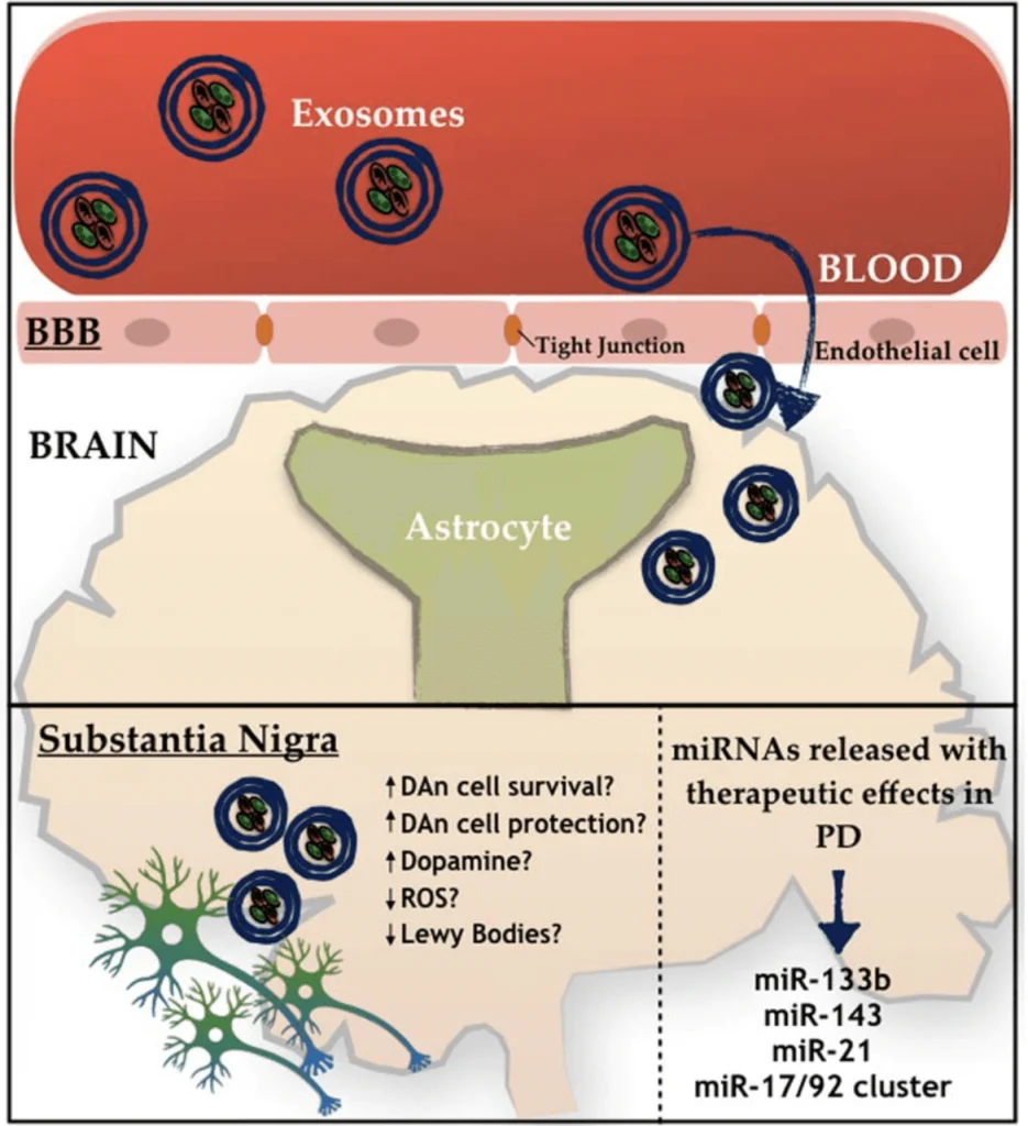
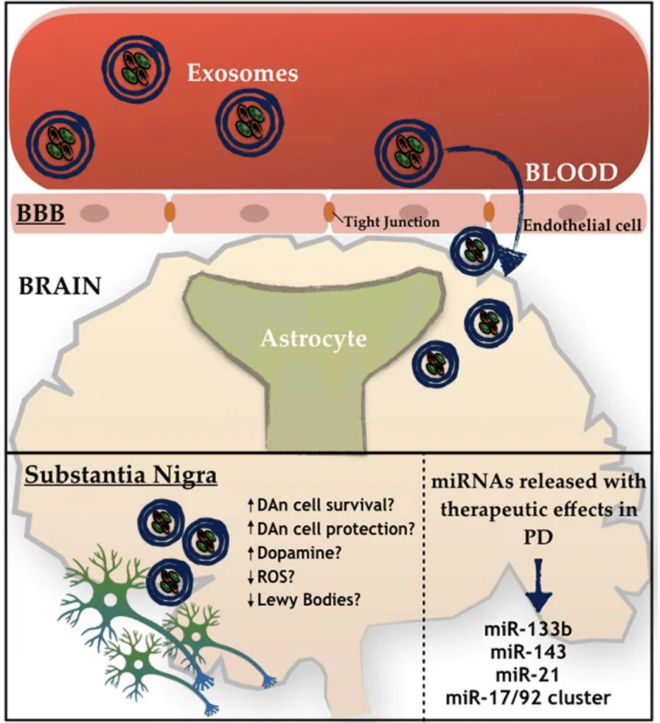
Recently, investigators suggested using Mesenchymal stem cells (MSCs)–derived exosomes as a therapy for different conditions, including Parkinson’s Disease (PD).[1,2,5] MSCs can be found in various body parts and specialize in different cell types depending on the body’s needs.[1] These cells produce extracellular vesicles called exosomes that have been studied as an alternative medicinal agent because of their stability and biological prospect in terms of the substances they carry, like signaling molecules, cytokines, enzymes, and micro-RNA (miRNA).[1,2,4] All these components are essential in maintaining cellular homeostasis, while the miRNA is more involved in regulating gene expression. [1,2,4] Many studies with MSCs have demonstrated several benefits in other neuropathological conditions. [1] One of the insights is that the MSCs have been pointed to activate different neuro-regeneration processes, opening a door for many possible ways to serve as promising therapies for future clinical trials. [1,3] Two targets for developing new treatments using MSCs are PD and Alzheimer’s disease (AD). [2,3,4,7] PD is characterized by the deterioration of dopaminergic neurons and the insufficiency of dopamine production. [3,6,9] Generally, the decrease of dopaminergic neurons is related to the accumulation of Lewy bodies (protein aggregates of α-synuclein) inside the neurons, which affects the normal functioning of those cells.[9] Interestingly, MSCs-derived exosome seems to be able to decrease one of the leading causes of PD, neuroinflammation.[2,5,10] On the other hand, AD is described as a brain illness that presents as neurological hallmarks the formation of amyloid plaques (Aβ) and neurofibrillary tangles causing synaptic loss.[4,7,12]

miRNA Researched for Parkinson’s Disease: Neuroprotection Blocks Neuron Death

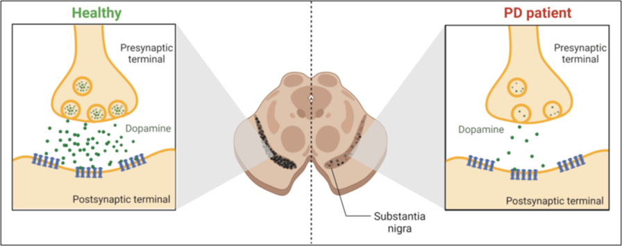
Programmed cell death, better known as apoptosis, is crucial in eliminating abnormal or unwanted cells in the body.[2] In recent years, the complex pathways regulating apoptosis have been well-studied as potential therapy targets.[2,10] Parkinson’s disease (PD) is no exception since the leading cause of this disorder is the death of dopamine-producing neurons in the brain (see Fig.1).[1-4] PD is a debilitating neurodegenerative disorder that steals a person’s ability to control movement.[8] Currently, levodopa (L-Dopa) is the most effective treatment for PD, helping in the improvement of the symptoms but not in the progression of the disease.[3] In addition, the use of L-Dopa led to adverse reactions after long-term administration.[3,7] Several studies propose using microRNAs (miRNAs) to inhibit the apoptosis of the dopamine-producing neurons found in PD.[6-9] miRNAs are tiny molecules capable of regulating gene expression (molecular switches) in the most critical processes for cell survival, like proliferation, cell differentiation, and apoptosis when required.[5-8]

Figure 1. The comparison between the normal functioning of dopaminergic neurons in healthy individuals vs in PD patients. 

For this reason, understanding the role of miRNAs in PD could be vital to developing new treatments to decrease the progression of the disorder. The apoptosis process is significant for eliminating unwanted cells.[2] Therefore, using miRNAs as a therapy has the potential to inhibit unwanted cell death and induce apoptosis in abnormal cells as well.[3-6] Understanding this interplay between miRNAs and apoptosis could lead to new treatment strategies for PD.[7] Several investigations found that miRNAs have neuroprotection abilities, safeguarding neurons from apoptotic cell death.[5] Alternatively, miRNAs promote apoptosis, thus eliminating damaged or dysfunctional neurons, which is crucial to clear cellular debris from microenvironments.[5]

Some benefits of using miRNAs as a therapy for PD are the following: (1) miRNAs can target specific genes involved directly in the apoptotic pathway (specificity). (2) miRNAs are molecular switches, activating or inhibiting the apoptosis pathway. This adaptive characteristic is very convenient for this type of therapy since PD requires, in some cases, the inhibition of neuronal death (dopamine-producing neurons) and the activation of apoptosis in unwanted cells in the brain. (3) miRNAs can cross the blood-brain barrier and enter into cells, which is a challenge in developing neurodegenerative diseases. The apoptosis pathway accelerates the progression of PD by the direct dopamine depletion caused by the death of dopamine-producing neurons.[4-6] As the disease progresses, more and more neurons are lost, leading to worsening symptoms and disability.

What is NAD+? How Does it Work?

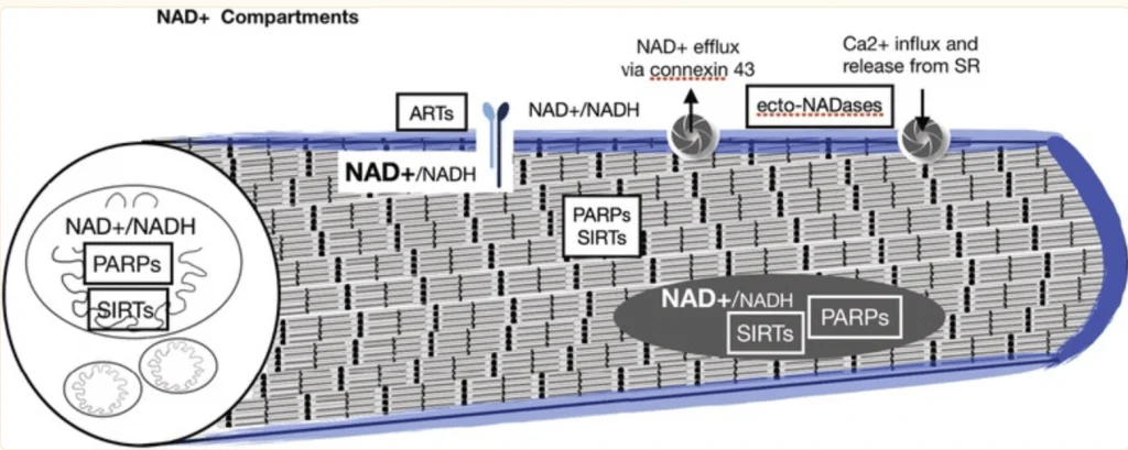
NAD+ is the second most abundant cofactor in the human body. Anti-aging therapies are becoming more mainstream as aging is now more often being viewed as a disease. Now that this transition is happening, the ability for NAD+ to activate PARPS, Sirtuins, and help with immune dysregulation has been thoroughly investigated and NAD+ and its precursors have been highly popularized. The clinical importance of maintaining cellular NAD+ levels was established early in the last century with the finding that pellagra, a disease characterized by diarrhea, dermatitis, dementia and death, could be cured with foods containing the NAD+ precursor niacin.

Additionally, cellular concentrations of NAD+ have been shown to decrease under conditions of increased oxidative damage such as occur during aging Altered levels of NAD+ have been found to accompany several disorders associated with increased oxidative/free radical damage including diabetes, heart disease, age-related vascular dysfunction, ischemic brain injury, misfolded neuronal proteins, and Alzheimer’s dementia. Interventions targeted at restoring NAD+ has been shown in animal models to support healthy aging and improve metabolic function, and dementia.

A need for NAD+ in muscle development, homeostasis, and aging

In a review study, researchers discuss the recent data that document conserved roles for NAD+ in skeletal muscle development, regeneration, aging, and disease as well as interventions targeting skeletal muscle and affecting NAD+ that suggest promising therapeutic benefits. The researchers also highlight gaps in our knowledge and propose avenues of future investigation to better understand why and how NAD+ regulates skeletal muscle biology.

How does 5-Amino-1MQ (and JBSNF) increase NAD+ and Metabolic Rate while Regenerating Aging Skeletal Muscle?

“Aging is accompanied by progressive declines in skeletal muscle mass and strength and impaired regenerative capacity, predisposing older adults to debilitating age-related muscle deteriorations and severe morbidity.

Muscle stem cells (muSCs) that proliferate, differentiate to fusion-competent myoblasts, and facilitate muscle regeneration are increasingly dysfunctional upon aging, impairing muscle recovery after injury. While regulators of muSC activity can offer novel therapeutics to improve recovery and reduce morbidity among aged adults, there are no known muSC regenerative small molecule therapeutics.

We recently developed small molecule inhibitors of nicotinamide N- methyltransferase (NNMT), an enzyme overexpressed with aging in skeletal muscles and linked to impairment of the NAD+ salvage pathway, dysregulated sirtuin 1 activity, and increased muSC senescence. We hypothesized that NNMT inhibitor (NNMTi) treatment will rescue age-related deficits in muSC activity to promote superior regeneration post-injury in aging muscle.

Results revealed that muscle stem cell proliferation and subsequent fusion were elevated in NNMTi-treated mice, supporting nearly 2-fold greater CSA and shifts in fiber size distribution to greater proportions of larger sized myofibers and fewer smaller sized fibers in NNMTi-treated mice compared to controls. Prolonged NNMTi treatment post-injury further augmented myofiber regeneration evinced by increasingly larger fiber CSA. Importantly, improved muSC activity translated not only to larger myofibers after injury but also to greater contractile function, with the peak torque of the TA increased by ~70% in NNMTi-treated mice compared to controls. Similar results were recapitulated in vitro with C2C12 myoblasts, where NNMTi treatment promoted and enhanced myoblast differentiation with supporting changes in the cellular NAD+/NADH redox states. Taken together, these results provide the first clear evidence that NNMT inhibitors constitute a viable pharmacological approach to enhance aged muscle regeneration by rescuing muSC function, supporting the development of NNMTi as novel mechanism-of-action therapeutic to improve skeletal muscle regenerative capacity and functional recovery after musculoskeletal injury in older adults.”

“NNMT inhibition using 5-amino-1MQ (30µM concentration) in both the pre-adipocytes (P < 0.01, treated pre-adipocytes vs. untreated controls) and the adipocytes (P < 0.05, treated adipocytes vs. untreated controls) resulted in significant reduction in the intracellular levels of 1-MNA…” (5)

Risk of Muscle Wasting in Elderly:

“The population of older (60+ years of age) adults is rapidly expanding in the United States and throughout the world, placing ever-increasing strains on health care resources and an urgent need for improved approaches to elder care. One of the most significant impacts of aging is the progressive decline in skeletal muscle mass and strength, with concomitant deteriorations in physical function and mobility that are strongly associated with numerous chronic diseases and increased mortality. While all older individuals experience muscle degeneration, approximately 30% of adults over 60 years of age and 50% of adults over 80 years of age develop sarcopenia, a geriatric disease characterized by significant and objective defects in muscle mass, strength, and function. Sarcopenic elderly individuals are at a 2-to 5-fold increased risk for permanent disability and greatly diminished quality of life arising from progressive muscle degeneration, decreased muscle function, and poor muscle quality that predispose them to debilitating falls and substantial disease burden. Furthermore, as muscle regenerative capacity of older adults becomes increasingly compromised, it leads to delayed and impaired recovery following muscle injury, decreased mobility and independence, increased hospitalization costs, and higher mortality rates.”
The Research Effects of TB-500 on Tissue Growth and Brain Injury

TB-500 is also known as thymosin beta 4 (TB4). Thymosin Beta 4 has been found, in animal models, to play a central role in controlling the structure of cells. By improving cell structure, TB-500 is thought to aid in wound healing, improve cell responses to stress, and even help cells to live longer. Scientific animal research studies have shown that TB-500’s role in regulating cell structure may eventually make it a leading therapeutic in wound healing, blood vessel repair, and even ocular (eye) repair.

Research has shown that when it comes to brain health, there are few drugs, supplements, or diets that make much difference. Unfortunately, the brain has remained a mystery to medical science and thus efforts to determine how to keep the brain healthy have been hindered. Science can tell us only that regular exercise and a relatively meat-free diet are associated with long-term brain health. There may, however, be some new evidence regarding thymosin beta 4 (also known as TB-4, or TB-500) and its impact on neural health.

Thymosin Beta 4

TB-500 (TB-4) is a naturally occurring peptide that is known to produce a vast array of healing and regenerative effects. It appears to promote everything from bone remodeling and growth after fracture to healing of heart muscle after a myocardial infarction (heart attack). Recent research in rats now suggests that TB-500 (TB-4) may improve neurological outcomes after stroke or brain damage.

What is NAD+? How Does it Work?

NAD+ is the second most abundant cofactor in the human body. Anti-aging therapies are becoming more mainstream as aging is now more often being viewed as a disease. Now that this transition is happening, the ability for NAD+ to activate PARPS, Sirtuins, and help with immune dysregulation has been thoroughly investigated and NAD+ and its precursors have been highly popularized. The clinical importance of maintaining cellular NAD+ levels was established early in the last century with the finding that pellagra, a disease characterized by diarrhea, dermatitis, dementia and death, could be cured with foods containing the NAD+ precursor niacin.

Additionally, cellular concentrations of NAD+ have been shown to decrease under conditions of increased oxidative damage such as occur during aging Altered levels of NAD+ have been found to accompany several disorders associated with increased oxidative/free radical damage including diabetes, heart disease, age-related vascular dysfunction, ischemic brain injury, misfolded neuronal proteins, and Alzheimer’s dementia. Interventions targeted at restoring NAD+ has been shown in animal models to support healthy aging and improve metabolic function, and dementia.

A need for NAD+ in muscle development, homeostasis, and aging

In a review study, researchers discuss the recent data that document conserved roles for NAD+ in skeletal muscle development, regeneration, aging, and disease as well as interventions targeting skeletal muscle and affecting NAD+ that suggest promising therapeutic benefits. The researchers also highlight gaps in our knowledge and propose avenues of future investigation to better understand why and how NAD+ regulates skeletal muscle biology.

Peptide Research for Neurocognitive Improvement.

Cognition is a complex system encompassing processes such as episodic memory, working memory, executive function/inhibition, spatial learning, language/vocabulary comprehension, processing speed, and language/reading decoding. Changes in synaptic plasticity, the ability of the brain to change and adapt to new information, can be short lived from milliseconds to years. Short lived forms include facilitation, augmentation, and potentiation which enhances neurotransmitter release.

These dynamic changes represent the molecular basis for learning and memory. This synaptic plasticity can be influenced by several factors e.g., aging, diseases (obesity, diabetes, hypertension, dyslipidemia), toxins (smoking and alcohol), and exercise. Aging has been estimated to trigger performance decline with an incidence of mild cognitive impairment of 21.5–71.3 per 1000 person-years). Cortical thickness and subcortical volume are shrinking 0.5–1% annually as a morphological sign of cognitive decline with plaques and axonal degeneration. Dementia is diagnosed when the acquired cognitive impairment has become severe enough to compromise social and/or occupational functioning with increasing prevalence.

Worldwide, around 50 million people have dementia and, with one new case every three seconds, the number of people with dementia is set to triple by 2050. Thus, there is a huge need for new research in order to combat the above-mentioned metrics. The peptides below have undergone extensive research to help aid in the improvement for our neurocognitive system.

Selank

Both Selank and Semax are melanocortin’s and have pleiotropic effects involved in brain health and function. Selank by itself has traditionally been prescribed for anxiety and depression. Selank has pronounced anxiolytic activity and acts as a stable neuropsychotropic, antidepressant, and anti-stress medication.

Semax

Semax is used as a therapeutic with pathologies related to brain circulation dysfunction. As a combination, Selank/Semax has applications in improving learning processes, exploratory behavior, regeneration and development, nociceptive and in amatory processes, accelerate nerve regeneration and improve neuromuscular performance and overall neural health.

Semax Research: Memory Enhancement and Brain Protection

Semax Stimulates Neurogenesis. 

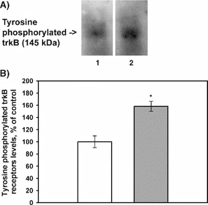
“Here, we found that a single application of Semax (50 μg/kg body weight) results in a maximal 1.4-fold increase of BDNF protein levels accompanying with 1.6-fold increase of trkB tyrosine phosporylation levels, and a 3-fold and a 2-fold increase of exon III BDNF and trkB mRNA levels, respectively, in the rat hippocampus. Semax-treated animals showed a distinct increase in the number of conditioned avoidance reactions. We suggest that Semax affects cognitive brain functions by modulating the expression and the activation of the hippocampal BDNF/trkB system.” (18)

Stem Cells Show Promise for Alzheimer’s and Parkinson’s Disease.

Mesenchymal stem cell-derived exosomes as a promising therapy for Parkinson’s and Alzheimer’s Disease.

Recently, investigators suggested using Mesenchymal stem cells (MSCs)–derived exosomes as a therapy for different conditions, including Parkinson’s Disease (PD).[1,2,5] MSCs can be found in various body parts and specialize in different cell types depending on the body’s needs.[1] These cells produce extracellular vesicles called exosomes that have been studied as an alternative medicinal agent because of their stability and biological prospect in terms of the substances they carry, like signaling molecules, cytokines, enzymes, and micro-RNA (miRNA).[1,2,4] All these components are essential in maintaining cellular homeostasis, while the miRNA is more involved in regulating gene expression. [1,2,4] Many studies with MSCs have demonstrated several benefits in other neuropathological conditions. [1] One of the insights is that the MSCs have been pointed to activate different neuro-regeneration processes, opening a door for many possible ways to serve as promising therapies for future clinical trials. [1,3] Two targets for developing new treatments using MSCs are PD and Alzheimer’s disease (AD). [2,3,4,7] PD is characterized by the deterioration of dopaminergic neurons and the insufficiency of dopamine production. [3,6,9] Generally, the decrease of dopaminergic neurons is related to the accumulation of Lewy bodies (protein aggregates of α-synuclein) inside the neurons, which affects the normal functioning of those cells.[9] Interestingly, MSCs-derived exosome seems to be able to decrease one of the leading causes of PD, neuroinflammation.[2,5,10] On the other hand, AD is described as a brain illness that presents as neurological hallmarks the formation of amyloid plaques (Aβ) and neurofibrillary tangles causing synaptic loss.[4,7,12]

Welcome back!

or
Menu
Explore

Search Peptides

Welcome back!

or
0